<?xml version='1.0' encoding='UTF-8'?>

<!DOCTYPE article PUBLIC "-//NLM//DTD JATS (Z39.96) Journal Publishing DTD v1.1d1 20130915//EN" "JATS-journalpublishing1.dtd">
<article xmlns:xlink="http://www.w3.org/1999/xlink">
  <front>
    <journal-meta id="journal-meta-87cddb9ab7774ac9973b6a64b7cbc767">
      <journal-id journal-id-type="nlm-ta">Sciresol</journal-id>
      <journal-id journal-id-type="publisher-id">Sciresol</journal-id>
      <journal-id journal-id-type="journal_submission_guidelines">https://jmsh.ac.in/</journal-id>
      <journal-title-group>
        <journal-title>Journal of Medical Sciences and Health</journal-title>
      </journal-title-group>
      <issn publication-format="print"/>
    </journal-meta>
    <article-meta>
        
          
            <article-id pub-id-type="doi">10.46347/jmsh.v12.i3.26.65</article-id>
          
          
            <article-categories>
              <subj-group>
                <subject>ORIGINAL ARTICLE</subject>
              </subj-group>
            </article-categories>
            <title-group>
              <article-title>&lt;p&gt;Cytological Detection and Seasonal Variation of Cervicovaginal Infections: A Two-Year Cross Sectional Observational Study from a Tertiary Care Centre&lt;/p&gt;</article-title>
            </title-group>
          
          
            <pub-date date-type="pub">
              <day>30</day>
              <month>3</month>
              <year>2026</year>
            </pub-date>
            <permissions>
              <copyright-year>2026</copyright-year>
            </permissions>
          
          
            <volume>12</volume>
          
          
            <issue>3</issue>
          
          <fpage>1</fpage>

          <abstract>
            <title>Abstract</title>
            &lt;p&gt;&lt;bold&gt;Background:&lt;/bold&gt; Cervicovaginal infections are common causes of morbidity among women of reproductive age. The major etiological agents include &lt;emphasis&gt;Bacterial vaginosis&lt;/emphasis&gt; (BV), &lt;emphasis&gt;Candida&lt;/emphasis&gt; species, and &lt;emphasis&gt;Trichomonas vaginalis&lt;/emphasis&gt; (TV). Although the Papanicolaou (Pap) smear is primarily used for cervical evaluation, it also serves as a practical tool for identifying infections and inflammatory conditions, especially in resource-limited settings where culture and molecular tests are not routinely available. &lt;bold&gt;Aims:&lt;/bold&gt;  Aim and objectives of the study are: 1) To determine the prevalence of BV, &lt;emphasis&gt;Candida&lt;/emphasis&gt;, and TV detected on conventional Pap smears; 2) To assess their seasonal variation.&lt;bold&gt; Methods: &lt;/bold&gt;A two-year cross-sectional study (January 2023–December 2024) was conducted at a tertiary care hospital in Udaipur, India. A total of 4860 satisfactory Pap smears from women undergoing routine gynecological screening were analyzed. Detection of BV, &lt;emphasis&gt;Candida&lt;/emphasis&gt;, and TV was based on cytomorphological features, and seasonal variation was evaluated across four defined Indian seasons. Statistical analysis was performed using the Chi-square test, with &lt;emphasis&gt;p&lt;/emphasis&gt; &amp;lt; 0.05 considered significant.&lt;bold&gt; Results: &lt;/bold&gt;Of 4860 smears, 1218 (25.1%) showed infections. BV was most frequent (58.8%), followed by &lt;emphasis&gt;Candida&lt;/emphasis&gt; (32.2%) and TV (9.0%). BV showed a significant seasonal peak during the monsoon (&lt;emphasis&gt;p&lt;/emphasis&gt; &amp;lt; 0.05), while &lt;emphasis&gt;Candida&lt;/emphasis&gt; and TV were distributed throughout the year.&lt;bold&gt; &lt;/bold&gt;BV is the predominant cervicovaginal infection detected on Pap smears, followed by &lt;emphasis&gt;Candida&lt;/emphasis&gt; and TV. &lt;bold&gt;Conclusion:&lt;/bold&gt; Seasonal surveillance may help identify temporal trends, enhance public health awareness, and guide empirical treatment strategies.&lt;/p&gt;
          </abstract>
          
          
            <kwd-group>
              <title>Keywords</title>
              
                <kwd>Pap smear</kwd>
              
                <kwd>&lt;I&gt;Candida&lt;/I&gt;</kwd>
              
                <kwd>&lt;I&gt;Trichomonas&lt;/I&gt;</kwd>
              
            </kwd-group>


        <contrib-group>
          
            
              <contrib contrib-type="author">
                <name>
                  <surname>Choudhary</surname>
                  <given-names>Ishita</given-names>
                </name>
                
                  <xref rid="aff-1" ref-type="aff">1</xref>
                
              </contrib>


              <aff id="aff-1">
                <institution> Senior Resident, Department of Pathology Dr. Sampurnanand Medical College </institution>
                <addr-line>Jodhpur India</addr-line>
              </aff>
            
              <aff id="aff-2">
                <institution> Associate Professor, Department of Pathology RNT Medical College </institution>
                <addr-line>Udaipur India</addr-line>
              </aff>
            
              <aff id="aff-3">
                <institution> Assistant Professor, Department of Pathology RNT Medical College </institution>
                <addr-line>Udaipur India</addr-line>
              </aff>
            
              <aff id="aff-4">
                <institution> Junior Resident, Department of Pathology RNT Medical College </institution>
                <addr-line>Udaipur India</addr-line>
              </aff>
            
              <aff id="aff-5">
                <institution> Senior Resident, Department of Pathology NSCB Medical College </institution>
                <addr-line>Jabalpur India</addr-line>
              </aff>
            
              <aff id="aff-6">
                <institution> Senior Resident, Department of Pediatrics RNT Medical College </institution>
                <addr-line>Udaipur India</addr-line>
              </aff>


              <contrib contrib-type="author">
                <name>
                  <surname>Kumari</surname>
                  <given-names>Priyanka</given-names>
                </name>
                
                  <xref rid="aff-2" ref-type="aff">2</xref>
                
              </contrib>


              <aff id="aff-1">
                <institution> Senior Resident, Department of Pathology Dr. Sampurnanand Medical College </institution>
                <addr-line>Jodhpur India</addr-line>
              </aff>
            
              <aff id="aff-2">
                <institution> Associate Professor, Department of Pathology RNT Medical College </institution>
                <addr-line>Udaipur India</addr-line>
              </aff>
            
              <aff id="aff-3">
                <institution> Assistant Professor, Department of Pathology RNT Medical College </institution>
                <addr-line>Udaipur India</addr-line>
              </aff>
            
              <aff id="aff-4">
                <institution> Junior Resident, Department of Pathology RNT Medical College </institution>
                <addr-line>Udaipur India</addr-line>
              </aff>
            
              <aff id="aff-5">
                <institution> Senior Resident, Department of Pathology NSCB Medical College </institution>
                <addr-line>Jabalpur India</addr-line>
              </aff>
            
              <aff id="aff-6">
                <institution> Senior Resident, Department of Pediatrics RNT Medical College </institution>
                <addr-line>Udaipur India</addr-line>
              </aff>


              <contrib contrib-type="author">
                <name>
                  <surname>Laddha</surname>
                  <given-names>Preeti</given-names>
                </name>
                
                  <xref rid="aff-3" ref-type="aff">3</xref>
                
              </contrib>


              <aff id="aff-1">
                <institution> Senior Resident, Department of Pathology Dr. Sampurnanand Medical College </institution>
                <addr-line>Jodhpur India</addr-line>
              </aff>
            
              <aff id="aff-2">
                <institution> Associate Professor, Department of Pathology RNT Medical College </institution>
                <addr-line>Udaipur India</addr-line>
              </aff>
            
              <aff id="aff-3">
                <institution> Assistant Professor, Department of Pathology RNT Medical College </institution>
                <addr-line>Udaipur India</addr-line>
              </aff>
            
              <aff id="aff-4">
                <institution> Junior Resident, Department of Pathology RNT Medical College </institution>
                <addr-line>Udaipur India</addr-line>
              </aff>
            
              <aff id="aff-5">
                <institution> Senior Resident, Department of Pathology NSCB Medical College </institution>
                <addr-line>Jabalpur India</addr-line>
              </aff>
            
              <aff id="aff-6">
                <institution> Senior Resident, Department of Pediatrics RNT Medical College </institution>
                <addr-line>Udaipur India</addr-line>
              </aff>


              <contrib contrib-type="author">
                <name>
                  <surname>Lodha</surname>
                  <given-names>Kritika</given-names>
                </name>
                
                  <xref rid="aff-4" ref-type="aff">4</xref>
                
              </contrib>


              <aff id="aff-1">
                <institution> Senior Resident, Department of Pathology Dr. Sampurnanand Medical College </institution>
                <addr-line>Jodhpur India</addr-line>
              </aff>
            
              <aff id="aff-2">
                <institution> Associate Professor, Department of Pathology RNT Medical College </institution>
                <addr-line>Udaipur India</addr-line>
              </aff>
            
              <aff id="aff-3">
                <institution> Assistant Professor, Department of Pathology RNT Medical College </institution>
                <addr-line>Udaipur India</addr-line>
              </aff>
            
              <aff id="aff-4">
                <institution> Junior Resident, Department of Pathology RNT Medical College </institution>
                <addr-line>Udaipur India</addr-line>
              </aff>
            
              <aff id="aff-5">
                <institution> Senior Resident, Department of Pathology NSCB Medical College </institution>
                <addr-line>Jabalpur India</addr-line>
              </aff>
            
              <aff id="aff-6">
                <institution> Senior Resident, Department of Pediatrics RNT Medical College </institution>
                <addr-line>Udaipur India</addr-line>
              </aff>


              <contrib contrib-type="author">
                <name>
                  <surname>Jain</surname>
                  <given-names>Minal</given-names>
                </name>
                
                  <xref rid="aff-5" ref-type="aff">5</xref>
                
              </contrib>


              <aff id="aff-1">
                <institution> Senior Resident, Department of Pathology Dr. Sampurnanand Medical College </institution>
                <addr-line>Jodhpur India</addr-line>
              </aff>
            
              <aff id="aff-2">
                <institution> Associate Professor, Department of Pathology RNT Medical College </institution>
                <addr-line>Udaipur India</addr-line>
              </aff>
            
              <aff id="aff-3">
                <institution> Assistant Professor, Department of Pathology RNT Medical College </institution>
                <addr-line>Udaipur India</addr-line>
              </aff>
            
              <aff id="aff-4">
                <institution> Junior Resident, Department of Pathology RNT Medical College </institution>
                <addr-line>Udaipur India</addr-line>
              </aff>
            
              <aff id="aff-5">
                <institution> Senior Resident, Department of Pathology NSCB Medical College </institution>
                <addr-line>Jabalpur India</addr-line>
              </aff>
            
              <aff id="aff-6">
                <institution> Senior Resident, Department of Pediatrics RNT Medical College </institution>
                <addr-line>Udaipur India</addr-line>
              </aff>


              <contrib contrib-type="author">
                <name>
                  <surname>Bisaria</surname>
                  <given-names>Anshuman</given-names>
                </name>
                
                  <xref rid="aff-6" ref-type="aff">6</xref>
                
              </contrib>


              <aff id="aff-1">
                <institution> Senior Resident, Department of Pathology Dr. Sampurnanand Medical College </institution>
                <addr-line>Jodhpur India</addr-line>
              </aff>
            
              <aff id="aff-2">
                <institution> Associate Professor, Department of Pathology RNT Medical College </institution>
                <addr-line>Udaipur India</addr-line>
              </aff>
            
              <aff id="aff-3">
                <institution> Assistant Professor, Department of Pathology RNT Medical College </institution>
                <addr-line>Udaipur India</addr-line>
              </aff>
            
              <aff id="aff-4">
                <institution> Junior Resident, Department of Pathology RNT Medical College </institution>
                <addr-line>Udaipur India</addr-line>
              </aff>
            
              <aff id="aff-5">
                <institution> Senior Resident, Department of Pathology NSCB Medical College </institution>
                <addr-line>Jabalpur India</addr-line>
              </aff>
            
              <aff id="aff-6">
                <institution> Senior Resident, Department of Pediatrics RNT Medical College </institution>
                <addr-line>Udaipur India</addr-line>
              </aff>
            
          
        </contrib-group>
        
    </article-meta>
  </front>
  <body>
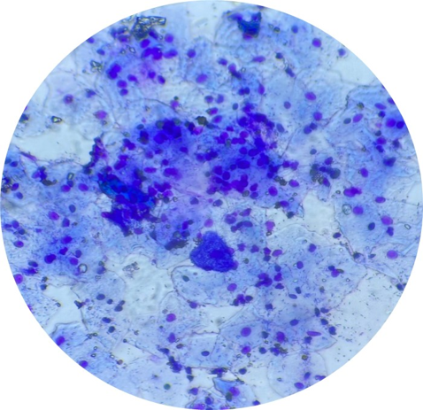
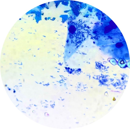
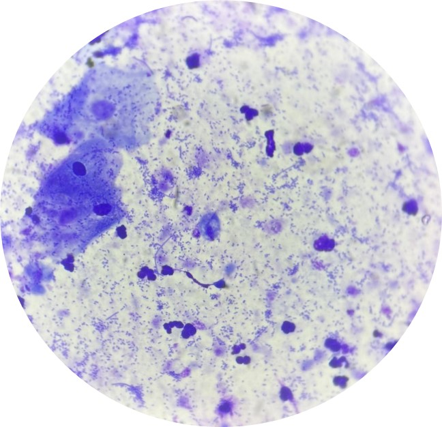
    <heading><span><bold>Introduction</bold></span></heading><p><span>Cervicovaginal infections are among the most common gynecological problems affecting women of reproductive age, contributing significantly to outpatient visits and adverse reproductive health outcomes<superscript>[<xref ref-type="link" rid="#ref-1">1</xref>]</superscript>. These infections are primarily caused by </span><italic><span>Bacterial vaginosis</span></italic><span> (BV), </span><italic><span>Candida</span></italic><span> species, and </span><italic><span>Trichomonas vaginalis</span></italic><span> (TV), each with distinct microbiological profiles and clinical presentations<superscript>[<xref ref-type="link" rid="#ref-2">2</xref>, <xref ref-type="link" rid="#ref-3">3</xref>]</superscript>.</span></p><div><span> </span></div><p><span>BV is characterized by an imbalance in the vaginal microflora, often associated with increased pH and clue cell formation; </span><italic><span>Candida</span></italic><span> is a fungal pathogen responsible for vulvovaginal candidiasis; and TV is a flagellated protozoan transmitted predominantly through sexual contact<superscript>[<xref ref-type="link" rid="#ref-4">4</xref>, <xref ref-type="link" rid="#ref-5">5</xref>]</superscript>.</span></p><p><span>The reported prevalence of these infections varies across India, with BV ranging from 15% to 45%, </span><italic><span>Candida</span></italic><span> from 3% to 30%, and TV from less than 1% to about 8%, depending on population characteristics, geographic region, and diagnostic modality<superscript>[<xref ref-type="link" rid="#ref-6">6</xref>, <xref ref-type="link" rid="#ref-7">7</xref>]</superscript>. In many resource-limited settings, culture and nucleic acid amplification tests (NAATs) are not routinely available, making cytological screening a valuable diagnostic adjunct.</span></p><p><span>The Papanicolaou (Pap) smear, while established as a cost-effective tool for cervical cancer screening, can also reveal cytological features of vaginal infections<superscript>[<xref ref-type="link" rid="#ref-8">8</xref>]</superscript>. </span></p><div><span> </span></div><p><span>This dual diagnostic role enhances its public health utility, particularly when linked to preventive strategies in primary care<superscript>[<xref ref-type="link" rid="#ref-9">9</xref>]</superscript>.</span></p><p><span>Given the potential influence of environmental and behavioral factors on infection rates, seasonal variation in cervicovaginal infections warrants systematic evaluation<superscript>[<xref ref-type="link" rid="#ref-10">10</xref>]</superscript>. Such patterns may reflect climatic effects on pathogen proliferation, host susceptibility, and health-seeking behavior, yet have not been widely studied in cytology-based datasets in India<superscript>[<xref ref-type="link" rid="#ref-11">11</xref>]</superscript>.</span></p><p><span>The present study aims to determine the prevalence of BV, </span><italic><span>Candida</span></italic><span>, and TV in women attending routine cervical cancer screening using conventional Pap smears, and to evaluate the seasonal distribution of these infections over a two-year period.</span></p><p><span><bold>Aim and Objectives:</bold></span></p><ordered-list><list-item><p><span>To determine the prevalence of BV, </span><italic><span>Candida</span></italic><span>, and TV detected on conventional Pap smears.</span></p></list-item><list-item><p><span>To assess their seasonal variation.</span></p></list-item></ordered-list><heading><span><bold>Material and Methods</bold></span></heading><p><span><bold>Study design</bold>: </span></p><p><span>This observational cross-sectional study was conducted at a tertiary care centre in Udaipur, India, over a two-year period from January 2023 to December 2024. </span></p><p><span><bold>Inclusion and exclusion criteria</bold>:</span></p><p><span>All satisfactory conventional Pap smears from women aged 18 years and above, submitted for routine gynecological screening during the study period, were included. Smears reported as unsatisfactory for evaluation, as per the Bethesda System 2014 criteria, were excluded.</span></p><p><span><bold>Sample size</bold>:</span></p><p><span>A total of 4860 satisfactory smears were included after exclusion of unsatisfactory cases.</span></p><p><span><bold>Data collection</bold>: </span></p><p><span>Patient demographic details (age), relevant clinical history (including symptoms such as vaginal discharge), and date of sample collection were retrieved from laboratory records. Information on intrauterine contraceptive device (IUCD) usage was not included in this analysis.</span></p><p><span><bold>Cytological evaluation</bold>: </span></p><p><span>The smears were immediately fixed in 95% ethyl alcohol for at least 15 minutes and stained using the conventional Papanicolaou staining technique. The procedure involved progressive nuclear staining with Harris hematoxylin, differentiation, bluing, and counterstaining with Orange G-6 (OG-6) and Eosin Azure (EA-36 or EA-50). The slides were then dehydrated, cleared in xylene, and mounted with DPX for microscopic examination. BV, </span><italic><span>Candida</span></italic><span>, and TV were identified based on standard cytomorphological criteria, as described in the Bethesda System. BV was diagnosed by the presence of clue cells, characterized by squamous epithelial cells with obscured borders due to adherent coccobacilli. </span><italic><span>Candida</span></italic><span> infection was identified by the presence of pseudohyphae and/or budding yeast forms. </span><italic><span>Trichomonas vaginalis</span></italic><span> was diagnosed by the identification of characteristic pear-shaped organisms with eccentrically placed nuclei and cyanophilic cytoplasm on Pap-stained smears Mixed infections were noted during evaluation but were excluded from statistical prevalence calculations in this study. A total of 762 mixed infection cases were identified and were excluded from pathogen-specific prevalence estimates to avoid double counting and ensure mutually exclusive comparisons across infection groups.</span></p><p><span><bold>Seasonal classification</bold>: </span></p><p><span>Cases were grouped into three Indian climatic seasons based on the date of sample collection:</span></p><p><span>Winter: December – February</span></p><p><span>Summer: March – May</span></p><p><span>Monsoon: June – November</span></p><p><span>The classification was based on regional climatic patterns of Southern Rajasthan, where monsoon-associated humidity commonly extends from June through November </span></p><p><span><bold>Statistical analysis</bold>: </span></p><p><span>Data were entered into Microsoft Excel and analyzed using SPSS version 20. Categorical variables were expressed as frequencies and percentages. The Chi-square test was used to compare infection prevalence across seasons. A p-value &lt;0.05 was considered statistically significant.</span></p><heading><span><bold>Results</bold></span></heading><p><span>A total of 4860 satisfactory conventional Pap smear samples collected over two years were included in the study. Among these, 1218 cases (25.1%) demonstrated cytological evidence of cervicovaginal infections.</span></p><p><span>The study population consisted predominantly of young and middle-aged adults. Individuals aged 20–29 years formed the largest group, accounting for 43.2% of cases, followed by those aged 30–39 years at 30.9%. Participants aged 40–49 years comprised 14.8% of the population, while the youngest (&lt;20 years) and oldest (≥50 years) age groups represented the smallest proportions, 6.2% and 4.9% respectively. Overall, over 88% of the study population fell between 20 and 49 years of age <xref ref-type="link" rid="#table-1">[Table. 1]</xref>.</span></p><figure id="table-1"><table><thead><tr><th><p><span><bold>Age Group (years)</bold></span></p></th><th><p><span><bold>Number of Cases</bold></span></p></th><th><p><span><bold>Percentage (%)</bold></span></p></th></tr></thead><tbody><tr><td><p><span>&lt;20</span></p></td><td><p><span>300</span></p></td><td><p><span>6.2%</span></p></td></tr><tr><td><p><span>20–29</span></p></td><td><p><span>2100</span></p></td><td><p><span>43.2%</span></p></td></tr><tr><td><p><span>30–39</span></p></td><td><p><span>1500</span></p></td><td><p><span>30.9%</span></p></td></tr><tr><td><p><span>40–49</span></p></td><td><p><span>720</span></p></td><td><p><span>14.8%</span></p></td></tr><tr><td><p><span>≥50</span></p></td><td><p><span>240</span></p></td><td><p><span>4.9%</span></p></td></tr></tbody></table><figcaption><span><bold>Table 1: Distribution of Study Population by Age Group (n=4860)</bold></span></figcaption></figure><p> </p><p><span>Bacterial vaginosis (BV) was the most frequently identified infection, found in 716 cases (58.8% of infections), followed by </span><italic><span>Candida</span></italic><span> species in 392 cases (32.2%), and </span><italic><span>Trichomonas vaginalis</span></italic><span> (TV) in 110 cases (9.0%) <xref ref-type="link" rid="#table-2">[Table. 2]</xref>.</span></p><figure id="table-2"><table><thead><tr><th><span><bold>Infection</bold></span></th><th><span><bold>Number of Positive Cases (n)</bold></span></th><th><span><bold>Percentage (%)</bold></span></th></tr></thead><tbody><tr><td><span>Bacterial Vaginosis (BV)</span></td><td><span>716</span></td><td><span>58.8%</span></td></tr><tr><td><italic><span>Candida</span></italic><span> species</span></td><td><span>392</span></td><td><span>32.2%</span></td></tr><tr><td><italic><span>Trichomonas vaginalis</span></italic><span> (TV)</span></td><td><span>110</span></td><td><span>9.0%</span></td></tr></tbody></table><figcaption><span><bold>Table 2: Prevalence of Cervicovaginal Infections Among Positive Cases (n=1218)</bold></span></figcaption></figure><p> </p><figure><graphic src="https://schoproductionportal.s3.ap-south-1.amazonaws.com/data/JMSH/337/1781869969745.png"/><figcaption><span><bold>Fig. 1: Pap smear showing clue cells characteristic of bacterial vaginosis (Pap stain, 100 x magnification)</bold></span></figcaption></figure><figure><graphic src="https://schoproductionportal.s3.ap-south-1.amazonaws.com/data/JMSH/337/1781869991832.png"/><figcaption><span><bold>Fig. 2: Pap smear showing pseudohyphae and budding yeast forms of </bold></span><italic><span><bold>Candida</bold></span></italic><span><bold> species (Pap stain, 100 x magnification)</bold></span></figcaption></figure><p> </p><figure><graphic src="https://schoproductionportal.s3.ap-south-1.amazonaws.com/data/JMSH/337/1781870011080.png"/><figcaption><span><bold>Fig. 3: Pap smear showing </bold></span><italic><span><bold>Trichomonas vaginalis</bold></span></italic><span><bold> organisms (Pap stain, 100 x magnification)</bold></span><o:wrapblock><graphic src="" uploadid="e0d32e098b331fe3d84508d36f726a8e5" uploadprocessed="true" v:shapes="Picture_x0020_3"/></o:wrapblock></figcaption></figure><p> </p><p><span>Seasonal distribution analysis demonstrated the highest detection rate of BV during the monsoon season (66.0%), compared to winter (51.3%) and summer (58.5%). </span><italic><span>Candida</span></italic><span> and TV infections showed less marked seasonal variation. The association between season and BV prevalence was statistically significant (Chi-square test, p &lt; 0.05), indicating a possible influence of climatic factors on the occurrence of bacterial vaginosis. No statistically significant seasonal variation was observed for </span><italic><span>Candida</span></italic><span> or TV infections (p &gt; 0.05) <xref ref-type="link" rid="#table-3">[Table. 3]</xref>.</span></p><figure id="table-3"><table><thead><tr><th><span><bold>Season</bold></span></th><th><span><bold>Total Infections</bold></span></th><th><span><bold>BV Cases (%)</bold></span></th><th><italic><span><bold>Candida  </bold></span></italic><span><bold>Cases (%)</bold></span></th><th><span><bold>TV Cases (%)</bold></span></th></tr></thead><tbody><tr><td><span>Winter</span></td><td><span>390</span></td><td><span>200 (51.3%)</span></td><td><span>130 (33.3%)</span></td><td><span>60 (15.4%)</span></td></tr><tr><td><span>Summer</span></td><td><span>410</span></td><td><span>240 (58.5%)</span></td><td><span>130 (31.7%)</span></td><td><span>40 (9.8%)</span></td></tr><tr><td><span>Monsoon</span></td><td><span>418</span></td><td><span>276 (66.0%)</span></td><td><span>132 (31.6%)</span></td><td><span>10 (2.4%)</span></td></tr></tbody></table><figcaption><span><bold>Table 3: Seasonal Distribution of Cervicovaginal Infections (n=1218)</bold></span></figcaption></figure><p> </p><heading><span><bold>Discussion</bold></span></heading><p><span>In the present study, out of 4860 satisfactory Pap smears, 25.1% showed evidence of infectious agents, with Bacterial vaginosis</span><italic><span> (BV)</span></italic><span> being the most common (58.8%), followed by </span><italic><span>Candida</span></italic><span> (32.2%) and </span><italic><span>Trichomonas vaginalis (TV)</span></italic><span> (9.0%).</span></p><p><span>Ranjit </span><italic><span>et al</span></italic><span>.<superscript>[<xref ref-type="link" rid="#ref-12">12</xref>]</superscript> found a 24.4% prevalence of BV among symptomatic women, consistent with our study. The significant associations with douching and abnormal discharge characteristics reaffirm known behavioral and clinical risk factors. Their observation that contraceptive use involving anatomical sites increased BV risk further supports the role of local vaginal environment disruption.</span></p><p><span>Disha </span><italic><span>et al</span></italic><span>.<superscript>[<xref ref-type="link" rid="#ref-13">13</xref>]</superscript> observed that </span><italic><span>Candida</span></italic><span> infections accounted for approximately 30% of vulvovaginal infections, a result that closely matches the 32.2% prevalence in the current study, further supporting the widespread occurrence of </span><italic><span>Candida</span></italic><span> as a significant pathogen in the female genital tract.</span></p><p><span>Vijaya </span><italic><span>et al.</span></italic><span><superscript>[<xref ref-type="link" rid="#ref-14">14</xref>]</superscript> reported a low prevalence of </span><italic><span>Trichomonas vaginalis</span></italic><span> infection in their study population. Given that the clinical presentation of trichomonal vaginitis often overlaps with other sexually transmitted infections, they emphasized the importance of laboratory confirmation for accurate diagnosis. According to their findings, both wet mount examination and culture remain effective and sensitive methods for detecting </span><italic><span>T. vaginalis</span></italic><span>.</span></p><p><span>Seasonal distribution analysis in the present study demonstrated the highest detection rate of BV during the monsoon season (66.0%), compared to winter (51.3%) and summer (58.5%). This pattern aligns with the findings of Sonti </span><italic><span>et al.</span></italic><span><superscript>[<xref ref-type="link" rid="#ref-5">5</xref>]</superscript>, who also reported increased detection of bacterial vaginosis during the rainy season, with BV identification reaching 24.9% in the monsoon compared with lower rates in winter (14.7%) and summer (11.8%). Their study similarly suggested that higher humidity and environmental moisture during the monsoon may promote alterations in vaginal microflora, predisposing women to BV. Together, these results reinforce the influence of seasonal climatic factors on BV prevalence and highlight the need for heightened clinical awareness during high-risk periods.</span></p><p><span>Overall, our findings corroborate previous Indian studies emphasizing BV as the most prevalent cervicovaginal infection, followed by </span><italic><span>Candida</span></italic><span> and </span><italic><span>Trichomonas vaginalis</span></italic><span>. The observed seasonal trend further highlights environmental and climatic influences on infection patterns, particularly for BV.</span></p><heading><span><bold>Conclusion</bold></span></heading><p><span>The present study highlights that cervicovaginal infections remain a significant finding on routine Pap smears, with </span><italic><span>Bacterial vaginosis</span></italic><span> emerging as the most prevalent, followed by </span><italic><span>Candida</span></italic><span> and </span><italic><span>Trichomonas vaginalis</span></italic><span>. The marked seasonal variation observed in BV, particularly its higher prevalence during the monsoon season, underscores the influence of environmental factors on infection dynamics. These findings emphasize the dual utility of Pap smears—not only as a tool for cervical cancer screening but also as a valuable method for detecting common infectious agents in low-resource settings. Incorporating seasonal surveillance into routine screening programs can aid in identifying temporal trends, enhancing public health awareness, and guiding timely preventive and therapeutic interventions.</span></p><p><span><bold>Limitations:</bold></span></p><p><span>The present study has certain limitations, being a single-center study, the findings may not be fully generalizable. In addition, diagnosis was based solely on cytomorphological findings without microbiological culture or molecular confirmation, which may affect diagnostic accuracy.</span></p>
  </body>
  <back>
    <ref-list>
      <title>References</title>
      
        
          <ref id="ref-1">
            <element-citation publication-type="journal">
              
                <person-group person-group-type="author">
                  <name>
                    Simões JA, Giraldo PC, Faúndes A.
                  </name>
                </person-group>
              
              
                <article-title>Prevalence of cervicovaginal infections during gestation and accuracy of clinical diagnosis</article-title>
              
              
                <source>Infectious Diseases in Obstetrics and Gynecology</source>
              
              
                <year>1998</year>
              
              
                <volume>6</volume>
              
              
                <issue>3</issue>
              
              
                <uri>https://doi.org/10.1002/(sici)1098-0997(1998)6:3&lt;129::aid-idog6&gt;3.0.co;2-s</uri>
              
            </element-citation>
          </ref>


          <ref id="ref-2">
            <element-citation publication-type="journal">
              
                <person-group person-group-type="author">
                  <name>
                    Sobel JD
                  </name>
                </person-group>
              
              
                <article-title>Vaginitis</article-title>
              
              
                <source>New England Journal of Medicine</source>
              
              
                <year>1997</year>
              
              
                <volume>337</volume>
              
              
                <issue>26</issue>
              
              
                <uri>https://doi.org/10.1056/nejm199712253372607</uri>
              
            </element-citation>
          </ref>


          <ref id="ref-3">
            <element-citation publication-type="journal">
              
                <person-group person-group-type="author">
                  <name>
                    Bhalla P, Chawla R, Garg S, Singh MM, Raina U, Bhalla R, &lt;I&gt;et al&lt;/I&gt;
                  </name>
                </person-group>
              
              
                <article-title>Prevalence of bacterial vaginosis among women in Delhi, India</article-title>
              
              
                <source>Indian Journal of Medical Research</source>
              
              
                <year>2007</year>
              
              
                <volume>125</volume>
              
              
                <issue>2</issue>
              
              
                <uri>https://doi.org/10.25259/IJMR_20071252_167</uri>
              
            </element-citation>
          </ref>


          <ref id="ref-4">
            <element-citation publication-type="journal">
              
                <person-group person-group-type="author">
                  <name>
                    Bisht M, Agarwal S, Upadhyay D
                  </name>
                </person-group>
              
              
                <article-title>Utility of Papanicolaou test in diagnosis of cervical lesions: a study in a tertiary care centre of western Uttar Pradesh</article-title>
              
              
                <source>International Journal of Research in Medical Sciences</source>
              
              
                <year>2015</year>
              
              
                <volume>3</volume>
              
              
                <issue>5</issue>
              
              
                <uri>https://doi.org/10.5455/2320-6012.ijrms20150508</uri>
              
            </element-citation>
          </ref>


          <ref id="ref-5">
            <element-citation publication-type="journal">
              
                <person-group person-group-type="author">
                  <name>
                    Sonti S, Sulochana S, Esakki Muthuvel E, Harikrishnan V, Srinivasan C
                  </name>
                </person-group>
              
              
                <article-title>Determination of prevalence and clinical significance of detection of microorganisms in Pap smear with seasonal variation</article-title>
              
              
                <source>National Journal of Laboratory Medicine</source>
              
              
                <year>2017</year>
              
              
                <volume>6</volume>
              
              
                <issue>3</issue>
              
              
                <uri>https://doi.org/10.7860/NJLM/2017/26474:2239</uri>
              
            </element-citation>
          </ref>


          <ref id="ref-6">
            <element-citation publication-type="journal">
              
                <person-group person-group-type="author">
                  <name>
                    Narasimha A, Nirup NC, Chandhana B, Nishanth N, Harendra Kumar ML
                  </name>
                </person-group>
              
              
                <article-title>Spectrum of infections in cervico-vaginal pap smears</article-title>
              
              
                <source>Journal of Clinical and Biomedical Sciences</source>
              
              
                <year>2014</year>
              
              
                <volume>04</volume>
              
              
                <issue>1</issue>
              
              
                <uri>https://doi.org/10.58739/jcbs/v04i1.4</uri>
              
            </element-citation>
          </ref>


          <ref id="ref-7">
            <element-citation publication-type="journal">
              
                <person-group person-group-type="author">
                  <name>
                    Leitich H, Bodner-Adler B, Brunbauer M, Kaider A, Egarter C, Husslein P
                  </name>
                </person-group>
              
              
                <article-title>Bacterial vaginosis as a risk factor for preterm delivery: A meta-analysis</article-title>
              
              
                <source>American Journal of Obstetrics and Gynecology</source>
              
              
                <year>2003</year>
              
              
                <volume>189</volume>
              
              
                <issue>1</issue>
              
              
                <uri>https://doi.org/10.1067/mob.2003.339</uri>
              
            </element-citation>
          </ref>


          <ref id="ref-8">
            <element-citation publication-type="journal">
              
                <person-group person-group-type="author">
                  <name>
                    Workowski KA, Bachmann LH, Chan PA, Johnston CM, Muzny CA, Park I, &lt;I&gt;et al&lt;/I&gt;
                  </name>
                </person-group>
              
              
                <article-title>Sexually Transmitted Infections Treatment Guidelines, 2021</article-title>
              
              
                <source>MMWR. Recommendations and Reports</source>
              
              
                <year>2021</year>
              
              
                <volume>70</volume>
              
              
                <issue>4</issue>
              
              
                <uri>https://doi.org/10.15585/mmwr.rr7004a1</uri>
              
            </element-citation>
          </ref>


          <ref id="ref-10">
            <element-citation publication-type="journal">
              
                <person-group person-group-type="author">
                  <name>
                    Crucitti T, Van Dyck E, Tehe A, Abdellati S, Vuylsteke B, Buve A, &lt;I&gt;et al&lt;/I&gt;
                  </name>
                </person-group>
              
              
                <article-title>Comparison of culture and different PCR assays for detection of Trichomonas vaginalis in self collected vaginal swab specimens</article-title>
              
              
                <source>Sexually Transmitted Infections</source>
              
              
                <year>2003</year>
              
              
                <volume>79</volume>
              
              
                <issue>5</issue>
              
              
                <uri>https://doi.org/10.1136/sti.79.5.393</uri>
              
            </element-citation>
          </ref>


          <ref id="ref-11">
            <element-citation publication-type="journal">
              
                <person-group person-group-type="author">
                  <name>
                    Fredricks DN, Fiedler TL, Marrazzo JM
                  </name>
                </person-group>
              
              
                <article-title>Molecular Identification of Bacteria Associated with Bacterial Vaginosis</article-title>
              
              
                <source>New England Journal of Medicine</source>
              
              
                <year>2005</year>
              
              
                <volume>353</volume>
              
              
                <issue>18</issue>
              
              
                <uri>https://doi.org/10.1056/nejmoa043802</uri>
              
            </element-citation>
          </ref>


          <ref id="ref-12">
            <element-citation publication-type="journal">
              
                <person-group person-group-type="author">
                  <name>
                    Ranjit E, Raghubanshi BR, Maskey S, Parajuli P
                  </name>
                </person-group>
              
              
                <article-title>Prevalence of Bacterial Vaginosis and Its Association with Risk Factors among Nonpregnant Women: A Hospital Based Study</article-title>
              
              
                <source>International Journal of Microbiology</source>
              
              
                <year>2018</year>
              
              
                <volume>2018</volume>


                <uri>https://doi.org/10.1155/2018/8349601</uri>
              
            </element-citation>
          </ref>


          <ref id="ref-13">
            <element-citation publication-type="journal">
              
                <person-group person-group-type="author">
                  <name>
                    Disha T, Haque F
                  </name>
                </person-group>
              
              
                <article-title>Prevalence and Risk Factors of Vulvovaginal Candidosis during Pregnancy: A Review</article-title>
              
              
                <source>Infectious Diseases in Obstetrics and Gynecology</source>
              
              
                <year>2022</year>
              
              
                <volume>2022</volume>


                <uri>https://doi.org/10.1155/2022/6195712</uri>
              
            </element-citation>
          </ref>


          <ref id="ref-14">
            <element-citation publication-type="journal">
              
                <person-group person-group-type="author">
                  <name>
                    Vijaya Mn D, Umashankar K, Sudha, Nagure AG, Kavitha G
                  </name>
                </person-group>
              
              
                <article-title>Prevalence of the &lt;I&gt;Trichomonas vaginalis&lt;/I&gt; Infection in A Tertiary Care Hospital in Rural Bangalore, Southern India</article-title>
              
              
                <source>Journal of Clinical and Diagnostic Research</source>
              
              
                <year>2013</year>
              
              
                <volume>7</volume>
              
              
                <issue>7</issue>
              
              
                <uri>https://doi.org/10.7860/jcdr/2013/5375.3140</uri>
              
            </element-citation>
          </ref>
        
      
    </ref-list>
  </back>
</article>