Journal of Medical Sciences and Health
DOI: 10.46347/jmsh.v12.i2.25.375
Year: 2026, Volume: 12, Issue: 2, Pages: 141-145
Original Article
P Rashmi 1, Barkha Rani 2, Akansh Khare 2, B M Soumya 3
1Assistant Professor of Pathology, Department of Pathology, J.J.M. Medical college, Davangere, Karnataka, India.
2Postgraduate, Department of Pathology, J.J.M. Medical college, Davangere, Karnataka, India.
3Associate Professor of Pathology, Department of Pathology, J.J.M. Medical college, Davangere, Karnataka, India.
Address for correspondence: Barkha Rani, Postgraduate, Department of Pathology, J.J.M. Medical college, Davangere, Karnataka, India.
E-mail: [email protected]
Received Date:11 December 2025, Accepted Date:07 April 2026, Published Date:18 May 2026

Introduction: Oral Squamous Cell Carcinoma (OSCC) continues to be a widely occurring cancer worldwide. Tumor-Associated Tissue Eosinophilia (TATE) refers to the infiltration of eosinophils within the tumor and the surrounding tumor-associated stroma. In OSCC, its prognostic significance is debated. Recent studies suggest that higher eosinophil density may correlate with better survival outcomes, while others link it with invasiveness.
Objectives: To evaluate the density of tissue eosinophils in OSCC and correlate their density with histopathological prognostic parameters.
Methods: A retrospective analysis carried out at the tertiary care center involving histopathologically confirmed cases of OSCC. Demographic, gross and microscopic features were recorded including quantification of tumor associated tissue eosinophils at 400x magnification on H&E-stained slides. The average eosinophil count per 10 high power fields (HPF) was determined, and their density was classified as negative, grade 1 (<10/HPF), grade 2 (11-20/HPF), and grade 3 (>20/HPF). It was compared with tumor grade, tumor size, depth of invasion, pattern of invasion and lymph node metastasis in available cases.
Results: The study involved 30 OSCC cases comprising 17 cases (56.6%) of well-differentiated OSCC, 10 cases (33.3%) of moderately differentiated OSCC, and 3 cases (1%) of poorly differentiated OSCC. The average age of the cases was 53 years, with the majority being male (21 cases). The site of the lesion was most commonly seen in buccal mucosa, followed by tongue, lip, alveolus and retromolar region. Most of the OSCC cases were ulcero-proliferative type with mean maximum dimension of 6.85cm. The TATE was observed in 24 instances (80%) and was negative in 6 instances (20%). The histological grade of OSCC was statistically significant with different grades of TATE (p = 0.037). Well differentiated OSCC showed greater levels of TATE when compared to PDSCC. Whereas no association was found between tumor size, depth of invasion, worst pattern of invasion and lymph node metastasis with different grades of TATE.
Conclusion: Elevated TATE is notably associated with well differentiated OSCC, suggesting that eosinophil infiltration may serve as a histological indicator of tumor differentiation. Future studies should include molecular profiling and clinical follow up to clarify TATE’s role in OSCC prognosis.
Oral Squamous Cell Carcinoma (OSCC) ranks as the sixth most prevalent cancer globally[6, 8, 9]. The incidence of OSCC is continually on rise because of various risk factors such as tobacco chewing, smoking, lack of nutrition, oro-dental factors, HPV infection etc.[1, 7].
Tumor Associated Tissue Eosinophilia (TATE) is defined as the presence of increased eosinophils infiltrating the tumor tissue and/or the tumor associated stroma at the primary tumor site, identified on histopathological examination of hematoxylin and eosin-stained sections[2, 4]. In Oral Squamous Cell Carcinoma (OSCC), the significance of its prognosis is a subject of debate. Recent studies suggest that higher eosinophil density may correlate with better survival outcomes, while others link it with invasiveness[1, 12].
A retrospective analysis was performed at the tertiary care center including cases of OSCC that were confirmed through histopathological examination. Cases were selected based on availability of adequate tumor tissue and complete clinicopathological data to minimize
potential confounding factors such as prior therapy or coexisting inflammatory conditions. Demographic data, gross and microscopic features were recorded including quantification of tumor associated tissue eosinophil count done at 400x magnification on H&E-stained slides.
Eosinophil counting was performed in areas showing the maximum density of eosinophilic infiltration (hotspot areas). Initially, the entire tumor section was scanned at low magnification (100×) to identify regions with the highest concentration of eosinophils. Subsequently, eosinophils were counted in 10 consecutive non-overlapping high-power fields (HPFs) at 400× magnification. The average eosinophil count per 10 HPFs was calculated for each case.
Based on eosinophil density TATE was graded as follows:
Negative 0 eosinophil
Grade 1 (<10 eosinophils /HPF)
Grade 2 (11-20 eosinophils /HPF)
Grade 3 (>20 eosinophils /HPF).
The eosinophil counts were correlated with tumor grade, tumor size, depth of invasion, pattern of invasion, and lymph node metastasis in the available cases.
Statistical analysis was performed using the Chi-square test to evaluate the association between TATE grades and clinicopathological parameters. A p-value <0.05 was considered statistically significant.
Individuals diagnosed with oral squamous cell carcinoma.
Individuals who have undergone wide excision and radical surgical procedures.
Individuals possessing complete clinicopathological data along with accessible tissue specimens.
Administration of preoperative chemotherapy or radiotherapy.
Insufficient tissue or poorly preserved samples.
Presence of coexisting inflammatory or allergic disorders.
The research encompassed 30 cases of OSCC, comprising 17 cases (56.6%) of well-differentiated OSCC, 10 cases (33.3%) of moderately differentiated OSCC, and 3 cases (10%) of poorly differentiated OSCC.
The mean age of the cases was 53 years (25y-75y) with majority of them being males (21 cases). The most common site of the lesion was buccal mucosa, followed by tongue, lip, alveolus and retromolar region.
Most of the OSCC cases were ulcero-proliferative type with mean maximum dimension of 6.85cm. The TATE was observed in 24 instances (80%) and was negative in 6 instances (20%). The histological grade of OSCC was statistically significant with different grades of TATE (p = 0.037) [Table. 1].
Well differentiated OSCC showed greater levels of TATE when compared to PD-SCC. Whereas no association was found between tumor size, depth of invasion, worst pattern of invasion and lymph node metastasis with different grades of TATE.
Among the patients, 22 had tobacco chewing habit, 3 alcoholics, 1 had both the habits and 4 patients had no such habits.
The most commonly involved site was buccal mucosa with other sites in descending order includes: Tongue>Lip>Alveolus/Retromolar trigone.
2 types of lesions were observed, ulceroproliferative and exophytic, with ulceroproliferative being more frequent.
| Tumor Grade |
TATE |
Total | |||
|---|---|---|---|---|---|
| Negative | Grade- 1 |
Grade- 2 |
Grade- 3 |
||
| WD | 1 | 3 | 8 | 5 | 17 (66.6%) |
| MD | 3 | 5 | 1 | 1 | 10 (27.1%) |
| PD | 2 | 1 | 0 | 0 | 3 (6.3%) |
| Total | 6 | 9 | 9 | 6 | 30 |
| CHI Square Test p value | 0.037 (Significant) | ||||
| Tumor Size |
TATE |
Total | |||
|---|---|---|---|---|---|
| Negative | Grade- 1 |
Grade- 2 |
Grade- 3 |
||
| <2 | 0 | 1 | 1 | 0 | 2 |
| 02-Apr | 2 | 5 | 0 | 1 | 8 |
| >4 | 4 | 3 | 8 | 5 | 20 |
| Total | 6 | 9 | 9 | 6 | 30 |
| CHI Square Test p value | 0.15 (Not Significant) | ||||
| Depth of Invasion |
TATE |
Total | |||
|---|---|---|---|---|---|
| Negative | Grade- 1 |
Grade- 2 |
Grade- 3 |
||
| ≤0.5 | 1 | 3 | 2 | 3 | 9 |
| 0.6-1 | 2 | 2 | 3 | 0 | 7 |
| >1 | 3 | 4 | 4 | 3 | 14 |
| Total | 6 | 9 | 9 | 6 | 30 |
| CHI Square Test p value | 0.74 (Not Significant) | ||||
| Pattern of Invasion |
TATE |
Total | |||
|---|---|---|---|---|---|
| Negative | Grade- 1 |
Grade- 2 |
Grade- 3 |
||
| 1 | 0 | 1 | 3 | 0 | 4 |
| 2 | 0 | 1 | 3 | 0 | 4 |
| 3 | 3 | 1 | 2 | 4 | 10 |
| 4 | 2 | 5 | 1 | 2 | 10 |
| WPOI-5 | 1 | 1 | 0 | 0 | 2 |
| Total | 6 | 9 | 9 | 6 | 30 |
| CHI Square Test p value | 0.58 (Not Significant) | ||||
| Lymph Node Mets |
TATE |
Total | |||
|---|---|---|---|---|---|
| Negative | Grade- 1 |
Grade- 2 |
Grade- 3 |
||
| Present | 3 | 2 | 1 | 3 | 9 |
| Absent | 3 | 7 | 8 | 3 | 21 |
| Total | 6 | 9 | 9 | 6 | 30 |
| CHI Square Test p value | 0.25 (Not Significant) | ||||
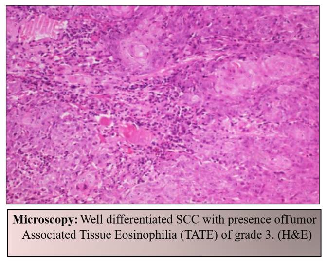
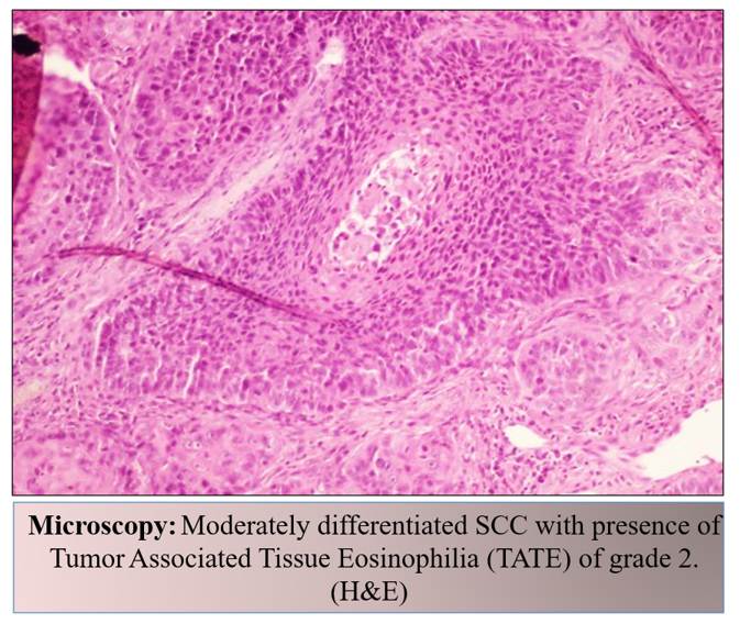
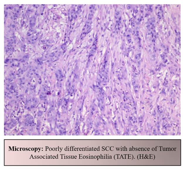
Eosinophils (granulocytic leukocytes) are myeloid precursors that are regulated by cytokines such as IL-3, granulocyte macrophage colony-stimulating factor, stem cell factor, IL-5, and various transcription factors. Mature Eosinophils, after circulating for 3 to 18 hours, migrate to the tissue, especially GIT, and under the influence of chemoattractants, they are recruited to the site of inflammation[10, 13].
TATE, also seen in other tumors like: TATE, in breast carcinoma (uncommon), is linked to high mutation or neoantigen load, but less cytologic immune activity[4, 5]. TATE, in colorectal carcinoma, eosinophils kills tumor cells directly and by anti-tumor immunity, linking to good prognosis[11].
In head and neck squamous cell carcinoma, TATE demonstrates a distinct behavior compared to other tumors, as eosinophils, which typically exhibit anti-tumor activity in other contexts, seem to play a tumor-promoting role in this case. This is primarily achieved through their induction of angiogenesis and metastasis[14].
Eosinophils demonstrate a dual role in tumor development and progression. On one hand, they exert anti-tumoral effects by releasing cytotoxic proteins such as eosinophil cationic protein and major basic protein, which can directly damage tumor cells. Additionally, eosinophils enhance tumor immunity by recruiting cytotoxic T-cells and natural killer cells. Conversely, eosinophils may also promote tumor progression through secretion of growth factors and pro-angiogenic mediators that facilitate tumor invasion and metastasis.
In many tumors, eosinophils support anti-tumor immunity by helping immune cells attack cancer, but under some conditions, they promote tumor growth[10]. Overall, TATE is associated with better survival, but not disease-free survival[3].
In this study, TATE was observed in 80% of the cases, with a significant association found between higher eosinophil counts and well-differentiated tumors (p = 0.037) This aligns with previous literature suggesting that eosinophilic infiltration may reflect a host immune response contributing to tumor suppression in well-differentiated carcinomas. The findings support the hypothesis that eosinophils may play a beneficial role in tumor immunity by releasing cytotoxic granules, chemokines, and cytokines that target tumor cells[12].
Similar findings were reported by Hosseinzadeh et al. (2024)[12], who observed that higher eosinophil infiltration was associated with well-differentiated OSCC, suggesting a potential role of eosinophils in tumor immune surveillance. Likewise, Choudhury et al. (2022)[7] demonstrated that tissue eosinophilia may reflect host immune response against tumor progression, although its association with metastatic potential remains inconsistent across studies.
Although higher grades of TATE were numerically more frequent in larger tumors (>4 cm), this association did not reach statistical significance (p = 0.15). This trend may reflect increased immune cell recruitment within larger tumor microenvironments, but studies with large number of cases are required to confirm this observation.
Moreover, eosinophils may participate in the recruitment and activation of other immune cells, enhancing the antitumor immune response[2]. Interestingly, no significant correlation was found between TATE and other prognostic parameters such as tumor size, depth of invasion, lymph node metastasis, or worst pattern of invasion[4]. This suggests that while TATE may reflect tumor differentiation, it may not be a reliable marker for tumor aggressiveness or metastatic potential. Discrepancies across studies regarding the prognostic role of TATE may stem from variations in sample size, methodology, and histological grading systems.
Elevated TATE is notably associated with well differentiated OSCC, suggesting eosinophil infiltration may serve as a histological indicator of tumor differentiation. Future studies should include molecular profiling and clinical follow up to clarify TATE’s role in OSCC prognosis.
Tumor-associated tissue eosinophilia (TATE) showed a significant association with well-differentiated OSCC in the present study, suggesting that eosinophilic infiltration may reflect host immune response and tumor differentiation. However, no significant association was observed with tumor size, depth of invasion, lymph node metastasis, or pattern of invasion. Larger prospective studies incorporating clinical outcomes and molecular profiling are required to further clarify the prognostic significance of TATE in OSCC.
1. Alshadwi A, et al. Eosinophilia as a prognostic marker in OSCC. Journal of Oral Pathology & Medicine. 2014;397–402.
2. Sahni P, Patel A, Md S, Hallur J, Gujjar PK. Tumor associated tissue eosinophilia in oral squamous cell carcinoma: A histo-chemical analysis. The Malaysian Malaysian Journal of Medical Sciences. 2015 Nov;22(6):21.
3. Tumor-associated Tissue Eosinophilia Predicts Favorable Clinical Outcome in Solid Tumors: A Meta-analysis. BMC cancer. 2020; 20 (1). Available from: https://doi.org/10.21203/rs.2.11177/v2
4. Ramu RS, et al. Role of eosinophils in the tumor microenvironment: Multifaceted roles in immunity and cancer. Immunologic Research. 2021;109–19.
5. Prevalence and clinical relevance of tumor-associated tissue eosinophilia (TATE) in breast cancer. Surgery. 2021; 169 (5). Available from: https://doi.org/10.1016/j.surg.2020.07.052
6. KMY YJ, et al. Eosinophil dynamics and tumor immunity in OSCC. Frontiers in Oncology. 2022;12:981345.
7. Choudhury S, et al. Evaluation of tissue eosinophilia with metastasis and recurrence in OSCC. Head and Neck Pathology. 2022;456–62.
8. Lu HY, et al. Tumor promoting and tumor-suppressing roles of eosinophils in cancer. Cancer Letters. 2023; 261–72.
9. Sinha S, et al. Eosinophils in OSCC and tumor progression. Cancer Investigation. 2023; 409–17.
10. Eosinophils in the tumor microenvironment: implications for cancer immunotherapy. Journal of Translational Medicine. 2023; 21 (1). Available from: https://doi.org/10.1186/s12967-023-04418-7
11. Tumor-associated tissue eosinophilia promotes angiogenesis and metastasis in head and neck squamous cell carcinoma. Neoplasia. 2023; 35 Available from: https://doi.org/10.1016/j.neo.2022.100855
12. Tumor-Associated Tissue Eosinophilia in Oral Squamous Cell Carcinoma: Implications for Histopathologic Grading. Archives of Razi Institute. 2024; 79 (6). Available from: https://doi.org/10.32592/ari.2024.79.6.1345
13. Eosinophils in Oral Disease: A Narrative Review. International Journal of Molecular Sciences. 2024; 25 (8). Available from: https://doi.org/10.3390/ijms25084373
14. Eosinophils in Colorectal Cancer: Emerging Insights into Anti-Tumoral Mechanisms and Clinical Implications. International Journal of Molecular Sciences. 2024; 25 (11). Available from: https://doi.org/10.3390/ijms25116098
Subscribe now for latest articles and news.