Journal of Medical Sciences and Health
DOI: 10.46347/jmsh.2016.v02i03.009
Year: 2016, Volume: 2, Issue: 3, Pages: 40-42
Case Report
Tanvi Meha1 , Anshul Raja2 , Anil Kumar Jaiswal1
1Department of Pediatrics, Patna Medical College & Hospital, Patna, Bihar, India,
2Department of Radiodiagnosis, Era’s Lucknow Medical College & Hospital, Lucknow, Uttar Pradesh, India
Address for correspondence:
Dr. Tanvi Meha, 403, Jaya Neelam Vihar, IAS Colony, Patna, Bihar, India. Phone: +91-9835490099.
E-mail: [email protected]
Williams–Campbell syndrome is a rare congenital syndrome characterized by the absence of cartilage in subsegmental bronchi leading to formation of bronchiectasis distal to the affected bronchi. The syndrome is usually presented in childhood, with recurrent pneumonia and broncho-obstructive symptoms such as coughing and wheezing. When patients’ signs and symptoms include recurrent respiratory infections and diffuse bronchiectasis, Williams–Campbell syndrome should be included in the differential diagnosis. The authors hereby report a case of a 1.5-year-old male child with respiratory distress who was diagnosed as Williams–Campbell syndrome on clinico-radiological grounds.
KEY WORDS:Pancreatitis, alcoholic, amylases, lipase, bilirubin
Williams–Campbell syndrome is a rare developmental disorder of familial occurrence which results due to the absence or deficiency of cartilage in the bronchial walls distal to first divisions of subsegmental bronchi and associated with diffuse cystic bronchiectasis. This uncommon entity should not be confused with congenital bronchiectasis which are those of hereditary conditions, such as cystic fibrosis, primary ciliary dyskinesia or immunodeficiency states that predispose to subsequent development of bronchiectasis.[1] The cartilage deficiency occurs in early life when the lungs are undergoing development. The exact mechanism is yet not understood. There is a lack of evidence suggesting any cartilage deficiency outside the lung. The trachea and central bronchi of the affected patients are of normal calibre. The symptoms and the prognosis thereof depend on the extent of involvement of cartilage maldevelopment in the bronchi.[2]
A 1.5-year-old male child presented to pediatrics emergency department with the complaints of fever, cough, and cold for 3 months and respiratory distress for 15days. The parents had consulted a local physician and were prescribed oral antibiotics, but the symptoms increased over the last 8 days and so the child was referred to a tertiary care center. The birth history was not eventful, the child was born by normal vaginal delivery at a government hospital and cried immediately after birth. There was no history of neonatal admission. He was immunized as per schedule and achieved all his milestones as per age. There was no history of any hospitalization before this event.
On general examination
The child was looking ill, had respiratory distress and was irritable at the time of admission. The child was febrile, respiratory rate - 70/min with use of accessory muscles of respiration, heart rate - 200/min, blood pressure - 90/62 mm of mercury, oxygen saturation on room air - 72%. Pallor was present along with significant bilateral cervical lymphadenopathy. On examination of respiratory system, there were diminished breath sounds in bilateral middle and lower zones with bilateral crepts. Examination of abdomen revealed hepatomegaly which was 6 cm below costal margin and was firm to hard in texture. Examination of cardiovascular system and central nervous system was normal.
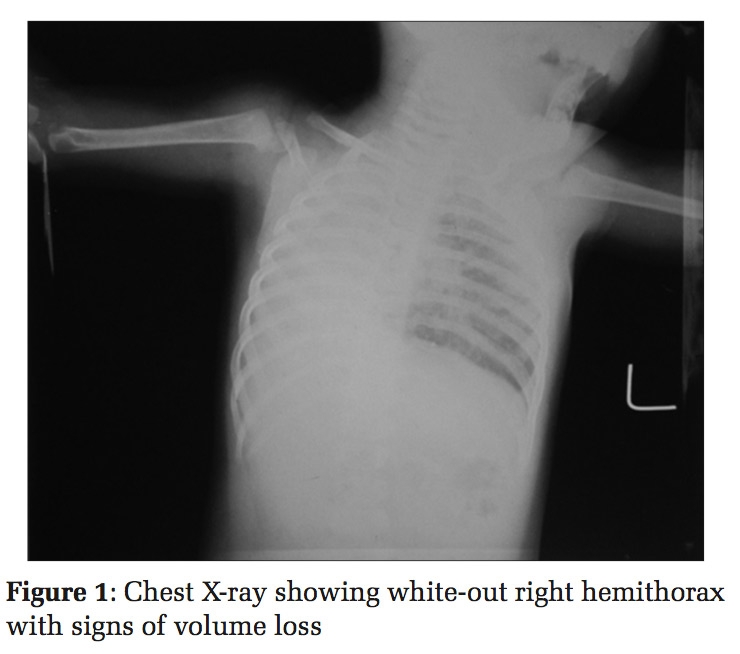
Laboratory investigations showed: hemoglobin - 10.3 g/dl, total count - 15,400/mm3, and platelet - 410,000/mm3. Renal function tests, serum electrolytes, routine examination of urine and stool, especially for the fat globules were normal. Liver function tests showed gamma-glutamyl transferase -843 U/L, serum glutamate-pyruvate transaminase - 91 U/L, and serum glutamic-pyruvic transaminase - 85 U/L. Culture of bronchoalveolar lavage was negative. Serum was negative for human immunodeficiency virus 1 and 2 antibodies. Serum immunoglobulin was normal. Antinuclear and antineutrophilic cytoplasmic antibody tests were negative. Serum alpha-1 antitrypsin level was normal. Aspergillus fumigatus specific immunoglobulin E was normal. Sweat test was negative. Chest roentgenogram revealed bilateral cystic shadows more prominent in the middle and lower zones. Ultrasonography of abdomen showed hepatomegaly with hypoechoic texture and mild ascites. Contrast-enhanced computed tomography showed bilateral hyperlucent lungs with cystic bronchiectasis. A diagnosis of cystic bronchiectasis due to Williams–Campbell syndrome was made based on clinico-radiological findings, laboratory testing, and medical history. Genetic testing was planned, but the patient refused further investigations due to financial constraints.
Williams–Campbell syndrome was originally described as a rare form of congenital bronchiectasis by Williams–Campbell et al. in 1960. They described a case series of five children who had similar clinical and radiological symptoms. It was postulated that the abnormal development of cartilage in bronchial tree was responsible for the disease.[3]
The diagnosis of Williams–Campbell syndrome requires an appropriate clinical history and characteristic expiratory collapse of airways after excluding other causes of congenital or acquired bronchiectasis. Earlier diagnosis relied on bronchography and fluoroscopic visualization, but these modalities did not offer good quality images. Fiberoptic bronchoscopy is often non- diagnostic and not all patients are good candidates for these procedures. During the 1990s, computed tomography became the radiological investigation of choice.[4] In 2006, Di Scioscio et al. described a case of Williams–Campbell syndrome using inspiratory and expiratory computed tomography imaging, which showed bilateral cylindrical/cystic bronchiectasis distal to the third-generation bronchi along with inspiratory ballooning and subsequent hyperinflation of the lung. There was a complete collapse of the bronchiectasis in the expiratory phase which pointed toward the absence of subsegmental bronchi cartilaginous plates.[5] George et al. on the same year used in addition to conventional computed tomography, an imaging technique termed the virtual bronchoscopy which used three-dimensional reconstruction of the bronchial tree. The case described by them showed collapsed subsegmental bronchi on conventional computed tomography; however on computed tomography bronchoscopy, the images were deficient of cartilage rings from the main stem to the subsegmental level.[6,7]
There is no specific treatment for Williams–Campbell syndrome. Prophylaxis from exacerbations is currently the recommended treatment. Prophylaxis is given until sputum production decreases which is achieved with an oral or intravenous antibiotic given for 7-10days. In severe cases, several antibiotics of different classes are used sequentially in a continuous regimen to prevent emergence of resistant strains. The treatment is tailored to meet the patient’s requirements and if warranted a bronchodilator combined with postural drainage and chest percussion can be used to remove secretions in a patient with bronchospasm and thick tenacious sputum.[8] Hypoxia warrants oxygen therapy. Non- invasive positive airway pressure can be used if respiratory acidosis persists.[9] Lung transplantation can be done when forced expiratory volume in 1 s (FEV1) reaches 30% of predicted value, when there is a rapid deterioration of FEV1, increase in frequency of exacerbations or exacerbation requiring intensive care unit stay, refractory/recurrent pneumothorax, and hemoptysis not controlled by artery embolization.[10,11] Williams–Campbell syndrome is an uncommon congenital cystic lung disease. The diagnosis of this syndrome is made by exclusion after detailed history, work up, and after ruling out the other common causes of congenital bronchiectasis.
Subscribe now for latest articles and news.