Journal of Medical Sciences and Health
DOI: 10.46347/jmsh.2017.v03i01.007
Year: 2017, Volume: 3, Issue: 1, Pages: 41-44
Case Report
Rekha Narendra Patil1, Satish Helwatkar2, Waman Raut3
Congenital granular cell epulis (CGCE) is a rare benign lesion of the newborn. It was first described in 1871 by Neumann. The exact etiology of the condition still remains unknown. There is marked female preponderance 10:1. The lesion is usually solitary, but multiple lesions are seen in 10% of cases. They arise from the gingival mucosa of the maxillary or mandibular alveolar ridge. Depending on the size of the lesion it can be asymptomatic or can cause feeding or respiratory problems. Imaging studies can help in the prenatal diagnosis of the lesion as early as 26 weeks of pregnancy. Spontaneous regression can occur, but surgery is the treatment of choice in symptomatic patients. CGCE is important as it has to be differentiated from the other aggressive lesions of early life. We report a case of 1 day old female neonate with intraoral lesion. The histopathology of the excised specimen helped in the diagnosis.
KEY WORDS:Epulis, gingival tumor, congenital granular cell epulis, neumann’s tumor, infant.
Congenital granular cell epulis (CGCE) is a rare benign congenital soft tissue growth of the newborn with an incidence of just 0.0006%. It was first described by Newmann in 1871 and hence also called as Neumann’s tumor.[1-12] There are many names for this condition such as CGCE, congenital epulis, congenital granular myoblastoma, congenital granular cell fibroblastoma[1,3-6,8,12] the WHO has recommended the term “CGCE” for this condition.[12]
Epulis is a Greek term meaning “of the gums.” Epulis is a nonspecific term used to describe variety of gingival lesions, regardless of their pathological origin.[1,4,8,12]
There is a marked female preponderance 10:1.[1,7,8,10,12]
It is mostly solitary, but multiple lesions are seen in 10% of cases with simultaneous involvement of both maxilla and mandible.[1,2,4,6-9,12] Other abnormalities such as nasal bridges, neurofibroma, polydactyly, Binder syndrome, congenital goiter can be seen with multiple CGCE.[8,12]
Depending on the size of the lesion it can be asymptomatic or can cause feeding and or respiratory problem.[1,2,4,7-9]
The recommended treatment is the surgical excision though spontaneous regression is known to occur.[1,2,6-10,12]
We present this case of 1 day old female neonate who had a gingival mass lesion on the maxillary alveolar ridge. The histopathological examination helped in diagnosing this rare condition. This is presented here to increase the awareness of this uncommon lesion.
This was 1 day old female neonate who was admitted with a mass lesion in the oral cavity. Relatives gave the history of difficulty in feeding. There was no history of respiratory distress. No history of fever.
The antenatal history of pregnancy was uneventful. There was no family history of hereditary diseases.
General physical examination did not show any obvious congenital abnormality apart from the gingival mass.
Routine laboratory investigations were within normal limits.
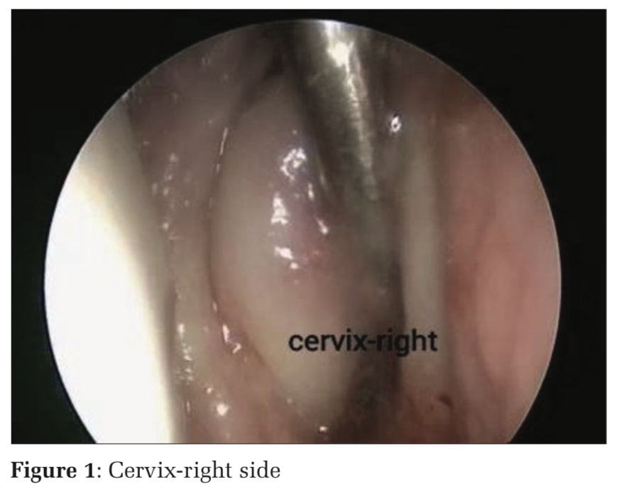
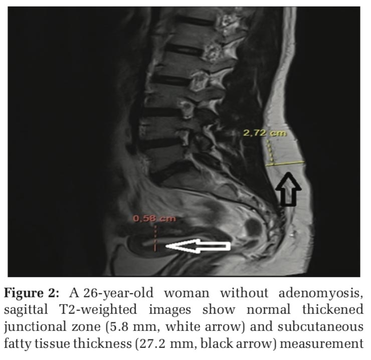
Local examination showed on oral mass of 2 cm × 2 cm. It was a pink colored, pedunculated mass arising from the maxillary gingiva. The color was similar to the color of the adjacent structures. It was firm nontender, noncompressible mass with smooth surface. Adjacent structures were normal.
After the proper discussion with the pediatrician, surgeon, and anesthetist; surgical excision was done. We received 2 cm × 2 cm single, firm, globular, pinkish-white tissue. The external surface was smooth, without any ulceration. Cut section was pinkish, homogenous without any areas of hemorrhage or necrosis.
Section showed stratified squamous epithelium. Subepithelial tissue showed closely packed sheets of cells having moderate to abundant amount of granular eosinophilic cytoplasm with round nuclei. There was scanty vascular stroma [Figures 1 and 2]. There was no nuclear pleomorphism, mitosis or necrosis. The stratified squamous epithelium did not show pseudoepitheliomatous hyperplasia.
Considering the clinical and microscopic findings the diagnosis of CGCE was given.


The CGCE is a rare oral mass lesion. The lesion is of concern for both parents and health-care professionals.[9] It is usually sporadic with no familial tendency.[8]
CGCE arises most commonly from the maxillary and mandibular alveolar ridges. The maxillary to mandibular involvement ratio being 3:1. This soft tissue lesion may have smooth pink mucosa which can be erythematous or ulcerated at times. It can be a sessile or pedunculated mass sometimes lobulated, size ranging from millimeters to few centimeters. Depending on the size it can be asymptomatic or can lead to feeding problems or respiratory distress.[1,4,6-9,12]
With the advances in the imaging technology, the prenatal diagnosis of the lesion can be possible as early as 26 weeks. Prenatal diagnosis is important, since large lesions may interfere with the vaginal delivery and a cesarean section can be planned. It can help in counseling the parents as to the nature of the condition and the treatment.[1,4,8,9,12]
The mass when large enough can cause oral obstruction which impairs the fetal deglutination and can result in polyhydramnios prenatally.[7,8,10]
The exact histogenesis is unclear. The proposed source of origin includes undifferentiated mesenchymal cells, odontogenic epithelial, pericytes, fibroblasts, smooth muscle cells. Trauma due to finger sucking in utero has also been suggested as one of the factors along with the degenerative or reactive hypothesis. Due to the high frequency seen in females an endogenous hormonal stimulus has also been considered, but this was unaccepted due to lack of detectable estrogen and progesterone receptors within the lesions.[1,2,4,7-10,12] Thediagnosisisusuallyclinicalandisconfirmedon histopathology.[9] As in the present case.
The growth stops at birth and spontaneous regression can occur. The treatment includes monitoring the patient for regression and in neonates with feeding or respiratory difficulties surgical excision can be done. Radical excision can damage the developing tooth buds and should be avoided. Malignant change and recurrence following incomplete excision has not been reported making wide local excision unnecessary.[1,8-10,12]
The histopathology shows a fairly circumscribed mass composed of nests and ribbons of tightly packed medium to large sized, polygonal to spindle cells with abundant granular eosinophilic cytoplasm. The nucleus is eccentric sometimes atypia can be seen with a small nucleoli, but with bland appearance. There is prominent capillary network. There is absence of rete ridges in the overlying stratified squamous epithelium.[5,12]
The lesion can also show angulated interstitial cells, occasional nests of odontogenic epithelium, cytoplasmic hyaline globules, lymphohistiocytic infiltrate and small peripheral nerve involvement and staghorn like vascular channels. Older and traumatic lesions show increased fibrosis and spindle cell features.[5,9,12]
Electron microscopy demonstrates intracellular granules (presumably autophagosomes), poorly formed junctional complexes and occasional long processes with contractile microfilaments.[12]
Histologically congenital granular cell tumor (CGCE) has to be differentiated from adult granular cell tumor. Adult granular cell tumor occurs in 30-60 years. It can also be seen in the tongue and other sites. The overlying stratified squamous epithelium shows the pseudoepitheliomatous hyperplasia.[1,3,8-10,12]
On immunohistochemistry the adult granular cell tumor shows positivity for S100, laminin, nerve growth factor receptor/p75, smooth muscle actin. CGCE (Congenital granular cell epulis) shows positivity for only vimentin, neuron specific enolase.[5,8,12]
The mass in the oral cavity can be seen in a variety of other conditions like congenital malformation such as encephalocele, dermoid cyst, teratoma and other benign and malignant neoplasm including vascular malformations, melanotic pigmented neuroectodermal tumor of infancy, and rhabdomyosarcoma. Melanotic pigmented neuroectodermal tumor is an osteolytic, pigmented infiltrating, neoplasm primarily affecting the jaws of the newborns. Predominance of female patient, tumor presence since birth, tumor location on the maxillary anterior region, and spontaneous regression without any intervention could rule out the majority of the differential diagnosis.[1,8,9,12]
In this case, the lesion was 2 cm × 2 cm, pedunculated mass arising from the maxillary gingival ridge. It had a smooth surface and was leading to feeding difficulty but no respiratory problem. The condition was not diagnosed on antenatal ultrasound check up. There was no hydramnios in the mother. This case was diagnosed after the histopathological examination of the excised specimen.
CGCE is a rare lesion present at birth mostly in female neonates. It is distressing to the parents and the health-care professionals. We provide the information about CGCE so that this condition can be more easily recognized and will increase the awareness of the lesion among the health-care givers. Although rare, the knowledge of this lesion is necessary for accurate diagnosis and proper treatment.
Acknowledgment The authors would like to thank the patient for the cooperation.
Subscribe now for latest articles and news.