Introduction
Appendicectomy consists of removing the entire appendix after dividing the mesoappendix and ligating the appendix base that connects to the cecum. Acute appendicitis is the most common surgical emergency and appendicectomy is routinely performed worldwide. The peak incidence of acute appendicitis is seen in teens and early 20s. It is more common in males than females 1. Around 20% of patients who undergo appendicectomy are found not to have acute appendicitis on histopathological examination 2.
Acute appendicitis is defined as inflammation of all the layers of the appendix. The exact pathogenesis of appendicitis is yet to be established 3, however luminal obstruction is considered the most common cause. The diagnosis of acute appendicitis is based on the patient’s history, laboratory and radiological findings, and subjective judgment based on his/her experience 4.
Histopathological examination of the appendix allows diagnosis of the pathological lesions as well as additional pathologies which may not be evident intra-operatively that can impact the patient’s management. This is more common in females than males. The diagnosis of acute appendicitis in many patients, especially in females, is difficult to
The practice of sending appendicectomy specimens for histopathological analysis varies suggest that appendices should not be sent routinely unless any gross abnormality is seen macroscopically in the appendix while operating 5. Histopathological examination remains the gold standard method for confirming it. It not only gives us the pathological diagnosis of acute inflammation, but the unusual finding of incidental tumors noted in the appendix, and it highlights the importance of the pathological analyses of every single resected appendix.
Materials and Methods
This cross sectional study was conducted in the Department of Pathology, National Institute of Medical Sciences and Research, Jaipur (Rajasthan). It was carried out for 2.5 years duration (January 2021- June 2023). It included 251 surgically removed either open or laparoscopic appendicectomy specimens sent for histolopathological evaluation to the Department of Pathology from National Institute of Medical Sciences and Research Hospital, Jaipur. Grossing was done and histopathologic sections was taken for the final diagnosis. Formalin-fixed paraffin-embedded sections was prepared and slides routinely stained with H&E.
With the use of SPSS software version 25, statistical analysis was performed on the findings related to age, clinical findings and histopathological findings. The proportion of neoplastic lesions was 3.58% comparing to prevalence rate in India of neoplastic cases (0.53%) and this is found highly statistically significant with p-value is 0.0000.
Results
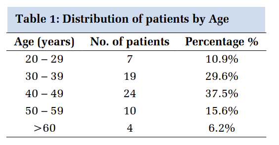
Out of the 251 cases, the majority of the cases were in the age group 20-30 years including male and female, accounting 54 cases (21.51%) followed by 47 cases (18.72%) in age group 30-40 years (Table 1). The mean age of the study population was 35.02 ± 16.72. Out of the total cases the majority were male 148(58.96%), while 103 (41.03%) were female. Only 09 (3.5%) cases were found of malignant aetiology on histopathological finding and shows statistically significant (p value-0.0000) as comparing with the prevalence rate of neoplastic cases in India i.e.0.53%. These three cases are of lymphoma, adenocarcinoma and carcinoid type of tumor. Male predominance was seen in most of the inflammatory lesions except 03 lesions i.e. Enterobius vermicularis (1.4%), Lymphoid Hyperplasia with Healing Appendicitis (0.71%) and acute appendicitis with vegetative material (0.71%). The inflammatory lesion that are found to be common in male are Lymphoid Hyperplasia (7.9%), Healing appendicitis (4.3%), Acute appendicitis (5%) and Acute on Chronic Appendicitis (2.14%). In neoplastic cases, male predominance is seen more accounting to 06 cases out of 9 cases (Figure 1) and in our study, majority of neoplastic cases were seen in young age group (20-40) years.
|
S.No. |
Age Interval (in years) |
Frequency |
Male |
Female |
|
1 |
0-10 |
11 |
07 |
03 |
|
2 |
10-20 |
43 |
25 |
18 |
|
3 |
20-30 |
54 |
31 |
23 |
|
4 |
30-40 |
47 |
29 |
18 |
|
5 |
40-50 |
42 |
23 |
19 |
|
6 |
50-60 |
29 |
16 |
13 |
|
7 |
60-70 |
16 |
10 |
06 |
|
8 |
70-80 |
9 |
07 |
02 |


Discussion
The histopathological examination of the appendix supports two things. First, it allows the diagnosis of acute appendicitis to be confirmed. Second, a histopathological examination of the appendix may reveal additional pathological findings that may not be seen on gross examination during operation. Among 251 study subjects in our study, the majority of them were in the age group 20-30 years i.e. 54 (21.51%) followed by 47 (18.72%) in 30-40 years. Similarly, the study done by Kulkarni MP (2017) 6 et al., reported 31.88% were in the age group 21-30 years. Other studies done by Shrestha R et. al. 7, Oguntola AS et. al. 8, V Vijayasaree et. al. 9 also revealed that around 80% of appendicitis occurred below forty years.
In our study, the majority (58.96%) of subjects were male while the females were 40.63%. Similar to the study findings of Zulfikar et al. (2009) 10, Sujatha R et. al. 11, Nabipour F (2003) 12 et al. and Duzgun AP (2004) 13 et al. reported the age range was wide with slight male preponderance.
Akbulut S et al. (2011) 14 have stated that obstruction is usually in the form of luminal obstructions such as fecolith, fibrosis, or stricture, which can lead to appendiceal gangrene and perforation. O’Connell PR (2010) 5 stated that obstruction can lead to the bacterial proliferation of aerobic and anaerobic organisms. However, in our study obstruction was seen in the form of fecolith, parasite and fibrosis. Sieren LM et al. (2010)15 revealed that a mucocele of the appendix denotes an obstructive dilatation of the appendiceal lumen due to abnormal accumulation of mucus, which may be caused either by a retention cyst, endometriosis, mucosal hyperplasia, cystadenoma, or a cystadenocarcinoma. The incidence of mucocele has been reported to range from 0.2% to 0.3% of all appendectomy specimens. In our study mucocele obstruction was not seen.
When assessing the distribution of signs & symptoms among the subjects, pain in the abdomen is the most frequent finding with (47.71%) of cases followed by Guarding (23.57%) of cases and Rigidity (20%). Vomiting was seen in 10 (7.14%) and fever in 5 (3.57%) cases. In Patel MM et al. (2007) 4 reported the most common presenting complaint was abdominal pain. This finding was concordant with other studies done by Sharma S et al. (2014) 16 and Edino et al. (2004) 17.
In daily practice, Koç C et al. (2019) 18 stated both neoplastic and nonneoplastic lesions of the appendix present with abdominal pain. Non-neoplastic lesions of the appendix outnumber the neoplastic counterparts, and appendicitis is the most frequently encountered lesion in daily clinical practice.
Acute appendicitis may be the mode of presentation of appendix neoplasms particularly adenocarcinoma as stated by Chan W & Fu KH (1987) 19. Although Patel M et. al. (2017) 4 revealed one case of adenocarcinoma and Primary appendiceal adenocarcinomas (PAAs) are very rare malignant neoplasms accounting for 0.05-0.2% of all appendectomies and only 6% of all malignant tumors of the appendix [Me O’ Donnell et al. (2007)] 20. But in our study, 03 cases of adenocarcinoma were reported accounting for 1.19%. In our study 05 case of Carcinoid type of tumor (1.99%) and one case of lymphoma (0.71%) was reported while Sieren LM et al. (2010) 15 stated that carcinoids are the most common tumors of the appendix and are typically small, firm, circumscribed yellow-brown lesions. An appendiceal carcinoid tumor is found in 0.3%- 2.27% of patients undergoing an appendectomy. In our study the proportion of neoplastic lesions was 3.58% comparing to prevalence rate in India of neoplastic cases (0.53%) and this is found highly statistically significant with p-value is 0.0000.
Conclusion
The present cross-sectional study was conducted on Surgically removed appendicectomy specimens and sent for histopathological evaluation showing incidence of appendicitis more in young age groups in males and this study also shows majority of neoplastic cases were seen in young age group. The objectives of study to analyse the incidence, age and gender distribution of operated cases of appendicectomy specimens and to evaluate the histopathological lesions (non-neoplastic and neoplastic) in appendicectomy specimens. Histopathological findings yield information about any neoplastic pathology present in cases with acute appendicitis like an unusual findings of carcinoid tumor, adenocarcinoma and b lymphoma. Therefore, this study concludes the necessity of histopathological examination for every appendicectomy specimens even in young individuals that mimics acute appendicitis.

