Journal of Medical Sciences and Health
DOI: 10.46347/jmsh.v10.i1.23.377
Year: 2024, Volume: 10, Issue: 1, Pages: 47-51
Original Article
G M Sreevidyalatha1 , P Shashikala2
1Assistant Professor, Department of Pathology, S.S. Institute of Medical Sciences and Research Centre, NH4, Bypass Road, Davangere, 577005,
2Professor & Head, Department of Pathology, S.S. Institute of Medical Sciences and Research Centre, NH4, Bypass Road, Davangere, 577005
Address for correspondence:
G M Sreevidyalatha, Assistant Professor, Department of Pathology, S.S. Institute of Medical Sciences and Research Centre, NH4, Bypass Road, Davangere, 577005.
E-mail: [email protected]
Received Date:10 November 2023, Accepted Date:02 February 2024, Published Date:08 April 2024
Background: Histopathology and Cytopathology are crucial aspects of medical education. With a paradigm shift from traditional to competency-based curriculum, teaching strategies are crucial to enable learning. we undertook a comparative analysis to evaluate the efficacy of two distinct teaching Learning tools—diagrams and microscopic images—in enhancing the observational and cognitive skills of medical students. Material and methods: A mixed method research design was planned to obtain quantitative data followed by qualitative data. These were later integrated for interpretation. A series of thirty microscopic images of cells, from topics already taught and diagrams of the same cells, drawn with Hematoxylin Eosin pencils, were projected to assess cell identification. Students were tasked with identifying and documenting their observations. The microscopic images were sourced from standard textbooks, while the diagrams were prepared by faculty and validated by subject experts. Responses were evaluated and scores analyzed using paired t-test. Focus group discussion was conducted to obtain qualitative data. Results: A group of 74 second-year medical students voluntarily participated in this study. Statistical analysis revealed that the scores for diagram identification were significantly higher than those for microscopic images, with a p-value of less than 0.05. Diagrams had a positive impact. Conclusion: Diagrams were superior to microscopic images in facilitating cell identification. This study underscores the importance of incorporating drawing-based teaching and learning methods in cytology as they encourage a more profound and effective learning process. Continued inclusion of diagrams in medical education to enhance students' cell identification skills is recommended.
Keywords: Cytopathology, Curriculum, Medical Education, Drawing
With a paradigm shift from traditional to competency-based curriculum and integrated teaching, devising suitable teaching strategies is crucial to enable learning and application of knowledge 1. Medical students are not pathologists or cytologists but they need to have a thorough knowledge of pathophysiology and pathology of various diseases which forms the basic foundation of medicine.
Pathology and its branches are an integral and crucial part of medical education and modular system-based curriculum. There is emphasis for teaching basic medical science with clinical relevance, thereby facilitating the application of basic knowledge in clinical context. Gross and microscopic appearances
There are only few studies to address the pedagogical methods in teaching cytopathology that facilitates application of knowledge and its clinical relevance.
The aim of this study is to optimize the teaching learning methods to enhance cell identification competency. The goal is to improve students’ ability to identify the cells by evaluating and comparing the impact of these two approaches-diagrams and microscopic images. By identifying the most effective method, the study aims to contribute to enriched learning experience, ensuring that students develop and refine their cell identification skills more efficiently and comprehensively. This enhancement in learning can ultimately lead to better prepared medical professionals with enhanced observational and cognitive skills.
To achieve the set goal, an explanatory sequential mixed method research method was designed. This consisted of initial quantitative data collection and statistical analysis, followed by qualitative phase to understand the impact of the preferred teaching learning tool. Later integration of both quantitative and qualitative data was done for complete interpretation.
Ethical clearance for the study was obtained from Institutes Ethical Clearance Board. The study included a group of second-year medical students who voluntarily participated in this study. The study was conducted in a classroom with conducive environment. This was not a part of their Formative assessment
To assess cell identification abilities, thirty microscopic images of cells (Set:1) and hand-drawn images of the same cells using Hematoxylin & Eosin (H&E) pencils (Set:2) were used. Microscopic images were projected first followed by Set 2 images in a different sequence. The cells included for the study were from topics already taught to them. Students were tasked with identifying and documenting their observations each time within a constant time-interval. The responses for both sets were collected and evaluated. Only the correct identification response was allotted 01 marks. The scores obtained by each student for both sets was tabulated and scores analyzed using paired t test
Focus group discussion was conducted to obtain the qualitative data of the learning tool using questionnaire, which was focused on eliciting their learning experiences, preference and the reasons for the same. The questionnaire was pilot tested. The students were divided into five groups by random sampling, with four groups of 15 students each and one group of 14 students. Consent was obtained, basic rules of discussion were communicated and confidentiality was assured to the participants. The discussion lasted for 60-80 minutes with a minimum of 12-15 minutes discussion on each question. The responses were recorded, transcribed and analyzed.
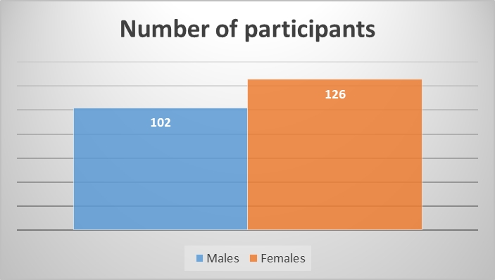
Seventy-four students willingly participated in the study. Male to female ratio was 1.5:1 with 44 male & 30 female students. The scores obtained for microscopic images ranged from 03 to 17, and 10 to 26 for H& E diagrams.

Statistical analysis revealed that the mean scores for H & E diagram identification (17.26 ± 2.73) were significantly higher than those for microscopic images (11.12 ± 3.19). Paired t-test showed a two-tailed p-value of less than 0.05 (Table 1).
|
|
Maximum
score |
Minimum
score |
Mean |
Standard
deviation |
P value |
|
Thirty Microscopic images (set 1) |
17 |
3 |
11.12 |
3.19 |
0.001 (<0.05) |
|
Thirty H & E diagram (set 2) |
26 |
10 |
17.26 |
2.73 |
Focus Group Discussion (FGD):responses were as follows.
Q1: Which set of images were easier to understand and identify?
The students inferred that H and E diagrams were easier to understand and identify the cells.
Q2: Which type of learning tool would you prefer in teaching learning methods and why?
Students opted for H & E diagrams as a teaching learning tool over microscopic image. Few reasons mentioned by students were” Diagrams give a clear picture to their imagination”, “It gives a three-dimensional perspective to understand the text as well as the microscopic images and has simplified the subject and the concepts”, “Cell identification and interpretation is not merely restricted to scoring marks in examinations but has a vast application in understanding the concepts”.
Q3: How did the preferred teaching tool enhance your learning?
Few responses given by students were “Diagrams are helpful in improving the academic performance as representative diagrams are self-explanatory, they convey and depict the same content instead of writing an entire paragraph”. “I need not byheart the concepts to remember, as drawings have helped me to develop long term memory and retain a pictorial memory of the topic”, “Diagrams made an imprint in memory and were easier to recollect & reproduce in exams”. “Using diagrams I can understand and analyze the concepts, learn complex changes occurring at cellular level leading on to the disease process-e.g.: dysplasia and metaplasia”
Q4: What were the draw backs of the other teaching learning tool?
Microscopic images were difficulty to identify. The reasons mentioned were” Microscopic images made it difficult in picking up the cell of interest which was supposed to be interpreted when found in relation to surrounding tissue”, “Microscopic images were difficult to recall and reproduce during assessments”. With regard to drawings the draw back cited was” I am not good at drawings and find it difficult and time consuming while using in tests and assessments”.
Q5. Take home message you inferred after being a part of the study.
Students felt that the study provided an insight to retrospect their study methods & identify their strengths and weakness. They were able to realize the importance of the concept of drawing in the regular teaching learning sessions and strong basic foundation which is very important to understand pathogenesis and clinical science”.
Overall qualitative data analysis predominantly (97.3%, n=72) revealed optimistic responses about diagrams. Diagrams were easy to appreciate & represent key points of structure, function & pathology. They were able to develop proper concepts and process data at cognitive level assessment Only two students (2.7%, n=2) expressed that drawings were complex and time consuming.
Pathology is not just seeing a slide! There is a need for relating what is seen to what is known about cytology and histopathology structure. Abraham Flexner emphasized almost seven decades back that the basic sciences should be taught with clinical relevance 2. Joewono in his studies has advocated and promoted the concept of drawing to learn in parallel with writing to learn 3. Studies have shown that Modern cytology and histopathology teaching methods using posters, digital photomicrographs and imaging fail to engage students with learning material 4, 5. It is found that there is lack of retention and clinical application among students 6. They are unable to completely and systematically comprehend cytology. It has only enabled students to see and read a slide but not remember or reproduce specific image.
In studies done by Weitman et al 7, Schwamborn et al8 & Kumar RK et al9 it was observed that drawings help students to acquire & remember information, perform well in tests. It enhanced deep learning and higher level of comprehension. Similar observations were made in Rafi et al 1. These observations were with the findings of our study. However, study done by Garcia M et al10 opined that students felt drawing is time consuming and is difficult for few who find it complex. This was the opinion in two students (2.7%) in our study. This can be addressed with more concentration on the basic structure rather than aesthetic or artistic ability.
Time immemorable, drawings are still considered to be an important teaching learning tool, where the learner engages in observation, comprehension & abstract conceptualization of structures under study 11. This teaching learning tool should not be compromised in an integrated medical curriculum with technological advancements.
Present study took students perspective into consideration as they are the important stakeholders of undergraduate teaching programme. Their opinion holds great significance in curriculum planning and implementation. There is a need to identify better educational strategies for the meaningful learning of cytology and histopathology.
Even regular lectures need to adapt similar drawing tools to simplify the subject. Teachers may have to spend more quality time and effort for preparing the diagrams 1. The data obtained is limited to II-year MBBS students. The study may be expanded to a larger cohort of students.
Diagrams were superior to microscopic images in facilitating cell identification. This study underscores the importance of incorporating drawing-based teaching and learning methods in cytology as they encourage a more profound and effective learning process. Continued inclusion of drawing based learning in medical education to enhance students cell identification skills is recommended.
Curriculum planners should encourage use of drawing as an important pedagogical tool as it uses psychomotor and cognitive skills for knowledge retention and understanding for its clinical application.
Subscribe now for latest articles and news.