Journal of Medical Sciences and Health
DOI: 10.46347/jmsh.v12.i3.26.103
Year: 2026, Volume: 12, Issue: 3, Pages: 347-350
Case Report
M Prishoo Reddy 1, Govindasamy Revathi 2, Erli Amel Ivan 3, V Sriram 4
1Post Graduate, Department of Pathology, Sri Manakula Vinayagar Medical college and Hospital, Puducherry-605107, India.
2Assistant Professor, Department of Pathology, Sri Manakula Vinayagar Medical College and Hospital, Puducherry-605107, India.
3Professor and Head, Department of Pathology, Sri Manakula Vinayagar Medical College and Hospital, Puducherry-605107, India.
4Professor, Department of Pathology, Sri Manakula Vinayagar Medical College and Hospital, Puducherry-605107, India.
Address for correspondence: Govindasamy Revathi, Assistant Professor, Department of Pathology, Sri Manakula Vinayagar Medical College and Hospital, Puducherry-605107, India.
E-mail: [email protected]
Received Date:06 March 2026, Accepted Date:29 June 2026, Published Date:19 August 2026

Hereditary elliptocytosis (HE) is an inherited red blood cell (RBC) membrane disorder characterized by elliptical or oval-shaped erythrocytes in peripheral smear. It is commonly inherited in an autosomal dominant pattern and results from defects in membrane skeletal proteins, particularly spectrin. Although many individuals are asymptomatic, some present with hemolytic anemia of varying severity. We report a case of hereditary elliptocytosis in a young female presenting with mild anemia, highlighting diagnostic challenges, importance of peripheral smear examination and family screening which remains crucial diagnostic tools in resource-limited settings.
Hereditary elliptocytosis (HE) is a heterogeneous group of inherited RBC membrane disorders characterized by the presence of elliptical or oval-shaped erythrocytes in peripheral blood[1]. This condition is most commonly inherited in an autosomal dominant pattern and results from mutations affecting proteins involved in maintaining erythrocyte cytoskeletal integrity, most commonly α-spectrin, β-spectrin, protein 4.1, and glycophorin C. The estimated global prevalence ranges from 1 in 2,000 to 1 in 4,000 individuals, with higher prevalence in malaria-endemic regions[2]. The criteria to diagnose HE includes presence of more than 25% of cells with elliptocyte morphology[3]. Clinical manifestations range from asymptomatic carrier states to severe hemolytic anemia, particularly in hereditary pyropoikilocytosis, a severe variant of HE. Diagnosis relies primarily on peripheral smear examination, supported by family history and molecular testing. Here, we report a case of Hereditary elliptocytosis in a young
female presenting with mild anemia, highlighting diagnostic challenges and the importance of peripheral smear examination and family screening.
A 16-year-old female presented to the General Medicine outpatient department with complaints of low-grade intermittent fever for five days, associated with chills and rigors, vomiting, abdominal pain for one day, and passage of clay-coloured stools for two days. There was no history of blood transfusion or significant bleeding episodes. The patient took native medication for three months. There was no significant family history of similar illness. On examination, patient had pallor, mild icterus, and moderate hepatomegaly.
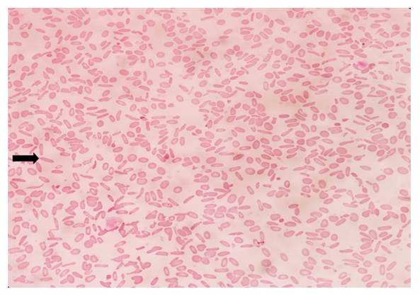
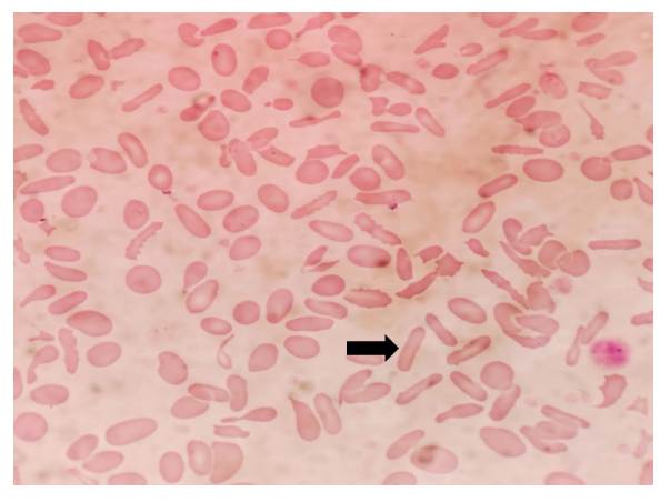
Initial laboratory investigations, including a complete blood count (CBC) revealed hemoglobin of 8.7 g/dL, packed cell volume of 28.7%, mean corpuscular volume (MCV) of 77.5 fL, mean corpuscular hemoglobin (MCH) of 28.9 pg, mean corpuscular hemoglobin concentration (MCHC) of 30.5 g/dL, total leukocyte count of 10,300 cells/mm³, and platelet count of 5.11 × 10⁵/mm³. Peripheral smear examination showed Microcytic hypochromic red cells admixed with few normocytes with moderate anisopoikilocytosis with approximately 60% elliptocytes, few tear-drop cells, occasional fragmented red cells, and polychromatophils. No spherocytes were observed [Fig. 1] & [Fig. 2]. The reticulocyte count was increased (7.5%). Both direct and indirect Coombs tests were negative. Sickling test and
osmotic fragility were also found to be negative. Serum lactate dehydrogenase was 221 IU/L. Liver function tests showed elevated indirect bilirubin, aspartate aminotransferase (AST), and alanine aminotransferase (ALT). Based on the peripheral smear findings, a provisional diagnosis of Microcytic hypochromic anemia with elliptocytosis (60%) was made.
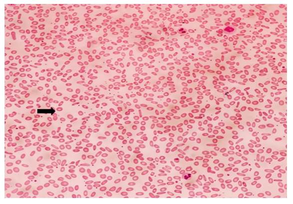
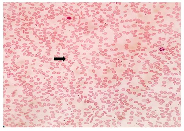
In view of a suspected inherited red cell membrane disorder, family screening was performed for the patient’s mother, father, and younger sister. Peripheral smear examination of the mother and younger sister revealed significant elliptocytosis (>50% and >40% elliptocytes), along with mild microcytic hypochromic anemia. Neither had a history of transfusion requirement or significant hemolytic episodes [Fig. 3] & [Fig. 4]. The father had normal hemoglobin levels and a normal peripheral smear. These findings strongly suggested an autosomal dominant pattern of inheritance. Based on the peripheral smear findings, laboratory parameters, and family screening, a diagnosis of Hereditary elliptocytosis was established.
Elliptocytosis was first described by Dresbach in the year 1904 and in 1932, Hunter first recognised it as a hereditary condition. It is inherited as an autosomal dominant trait except for hemolytic HE and Hereditary pyropoikilocytosis, where the inheritance is autosomal recessive. The incidence of hereditary elliptocytosis is difficult to determine because more than 90% of the people with elliptocytosis are asymptomatic[1]. Both sexes are equally affected. HE is common in people of Africa and Mediterranean region[4]. Since elliptocytosis are resistant to malaria, some subtypes of elliptocytosis are more prevalent in areas where malaria is endemic[5]. HE is usually discovered as an incidental finding during peripheral blood smear examination, but most consistent and characteristic smear findings in all variants of HE is the presence of more than 25 % elliptocytes on PS and usually more than 60%[6].
It occurs due to disruption of horizontal protein-protein interaction i.e. defect in self association of αß spectrin heterodimer to form tetramer (most common) and defect in junctional complex protein–protein interaction (Protein 4.1R, Glycophorin C). This membrane instability leads to alteration in the membrane deformability and mechanical properties leading to elliptocytosis[5, 7]. Approximately 65% of cases of hereditary elliptocytosis have mutations of alpha spectrin, 30% beta spectrin, and 5% protein 4.
Hereditary Elliptocytosis syndromes are classified into several groups includes hereditary elliptocytosis (HE), hemolytic hereditary elliptocytosis, hereditary pyropiokilocytosis (HPP), spherocytic hereditary elliptocytosis and South East Asian Ovalocytosis (SAO). They are characterized by clinical, biochemical and genetic heterogeneity. Most patients are clinically asymptomatic, and the diagnosis is usually made incidentally when a peripheral blood smear is examined. Asymptomatic patients are heterozygous for the disease and have mild anemia or vague symptoms like abdominal pain as in our case. Approximately, 10% of patients have moderate to severe anemia, with intermittent episodes of acute hemolysis with jaundice and splenomegaly. Our case had mild microcytic hypochromic anemia and jaundice without evidence of acute hemolysis. The reason for microcytosis can be due to coexisting iron deficiency state. Iron studies was not done during the evaluation and therefore the exact cause of the microcytic anemia could not be established.
The differential diagnosis considered are Iron deficiency anemia because of the microcytic hypochromic red cell indices. However, isolated iron deficiency is unlikely to account for the marked elliptocytosis observed in our patient and affected family members. Hereditary spherocytosis was excluded by the absence of spherocytes on peripheral smear and normal osmotic fragility testing. Autoimmune hemolytic anemia was ruled out by negative direct and indirect antiglobulin (Coombs) tests. Thalassemia trait was also considered because of the microcytic indices, but the peripheral smear lacked characteristic target cells and the familial occurrence of marked elliptocytosis strongly supported HE. Acquired elliptocytosis associated conditions were less likely because of the age of presentation, clinical features, and demonstration of similar peripheral findings in first-degree relatives.
Diagnosis of hereditary elliptocytosis mainly depends on morphology of red cells, mainly elliptical red blood cells on peripheral blood smear, and eosin -5-maleinide (EMA) binding method[8, 9]. But we made diagnosis with RBC morphology in the peripheral smear.
On further evaluation, family screening was performed in view of the suspected inherited red cell membrane disorder. Peripheral smear examination of the patient’s mother and younger sister revealed significant elliptocytosis (>30% elliptocytes) with mild anemia in the mother and sister and normal hemoglobin levels in the father. These findings strongly support an autosomal dominant inheritance pattern, which is the most common mode of transmission in hereditary elliptocytosis (HE).
Identification of affected but asymptomatic family members underscore the variable clinical presentation of HE. Hence, genetic counselling should be given to the family members regarding inheritance risk, especially in reproductive-age individuals. Early recognition allows appropriate counselling and prevents unnecessary investigations and therapeutic interventions.
Conservative management with folate supplementation and observation is sufficient in mild cases. In severely anemic patients, transfusion may be needed. Splenectomy is indicated for severe form of elliptocytosis after 5 years of age which increases the red cell life span and haemoglobin levels[10].
The diagnostic difficulties in this case was that patient presented with fever, jaundice, hepatomegaly, and showed microcytic hypochromic anemia rather than with classical manifestations of a hereditary red cell membrane disorder. The reduced hemoglobin, MCV and MCH values initially raised the possibility of iron deficiency anemia. Furthermore, the coexistence of microcytosis and hypochromia obscured the diagnostic significance of elliptocytosis, particularly in the absence of a known family history of hereditary elliptocytosis. But, the diagnosis was made through careful peripheral smear examination, which showed approximately 60% elliptocytes, supported by evidence of reticulocytosis and the identification of similar findings in the first-degree relatives. The limitation of this case report was the confirmatory tests like Eosin-5-Maleimide (EMA) binding assay and molecular genetic testing were not done.
This case highlights the enduring diagnostic value of peripheral smear examination and family screening, especially in resource-limited settings where specialized red cell membrane studies and molecular testing were readily unavailable. This case report is significant because it illustrates how hereditary elliptocytosis can remain clinically undiagnosed until adolescence and may be masked by coexisting microcytic anemia and nonspecific clinical features, thereby delaying diagnosis.
1. Hoffman R, Benz EJ Jr, Shattil SJ, Furie B, Cohen HJ, Silberstein LE, et al. Red cell membrane disorders. Red cell membrane disorders. In: Hematology Basic Principles and Practice. Philadelphia USA. Churchill Livingstone; 2000. 576- 610.
2. Abnormal Red Blood Cell Morphology in an Elderly Male. Laboratory Medicine. 2004; 35 (9). Available from: https://doi.org/10.1309/k346-5cr3-7p6y-24g5
3. Bain B. Red cell membrane disorders. In: Blood Cells: a practical guide. 4th ed. Wiley Blackwell; 2006. p. 278-281.
4. Southeast Asian ovalocytosis in an African-American family [letter]. Blood. 1994; 84 (8). Available from: https://doi.org/10.1182/blood.v84.8.2823.2823
5. Hereditary elliptocytosis: spectrin and protein 4.1R. Seminars in Hematology. 2004; 41 (2). Available from: https://doi.org/10.1053/j.seminhematol.2004.01.003
6. Hereditary Elliptocytosis and Hyperhaemolysis. Acta Medica Scandinavica. 1966; 179 (6). Available from: https://doi.org/10.1111/j.0954-6820.1966.tb05496.x
7. The molecular basis of disorders of the red cell membrane.. Journal of Clinical Pathology. 1999; 52 (4). Available from: https://doi.org/10.1136/jcp.52.4.245
8. Guidelines for the diagnosis and management of hereditary spherocytosis. British Journal of Haematology. 2004; 126 (4). Available from: https://doi.org/10.1111/j.1365-2141.2004.05052.x
9. Rapid flow cytometric test for the diagnosis of membrane cytoskeleton-associated haemolytic anaemia. British Journal of Haematology. 2000; 111 (3). Available from: https://doi.org/10.1046/j.1365-2141.2000.02416.x
10. Hereditary spherocytosis, elliptocytosis, and other red cell membrane disorders. Blood Reviews. 2013; 27 (4). Available from: https://doi.org/10.1016/j.blre.2013.04.003
Subscribe now for latest articles and news.