

Journal of Medical Sciences and Health
DOI: 10.46347/jmsh.2018.v04i02.003
Year: 2018, Volume: 4, Issue: 2, Pages: 18-21
Case Report
Ipsita Acharya, Sanjib Kumar Das, Jayashree Mohanty, Sasmita Parida
Departement of Radiodiagnosis, Srirama Chandra Bhanja Medical College, Cuttack – 753 007, Odisha, India
Address for correspondence:
Dr. Sanjib Kumar Das, Department of Radiodiagnosis, Srirama Chandra Bhanja Medical College, Cuttack – 753 007, Odisha, India. Phone: +91-9861463215. E-mail: [email protected]
Retrocaval ureter or circumcaval ureter is a rare developmental anomaly of inferior vena cava (IVC) where the ureter lies posterior to IVC, again to cross anteriorly below. Right ureter is most commonly though not exclusively involved. It is most commonly asymptomatic, but sometimes it may lead to stasis owing to obstruction of ureter which leads to hydronephrosis with proximal hydroureter. We present a case of retrocaval ureter in a young female patient presenting with recurrent right flank pain.
KEY WORDS:Circumcaval ureter, hydroureteronephrosis, recurrent flank pain.
Retrocaval ureter is an uncommon cause of hydroureteronephrosis in young adults. Although generally not suspected on ultrasonography (USG), contrast-enhanced computed tomography (CECT)/ magnetic resonance urography (MRU) can readily diagnose this condition facilitating early detection and proper management.
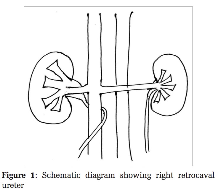
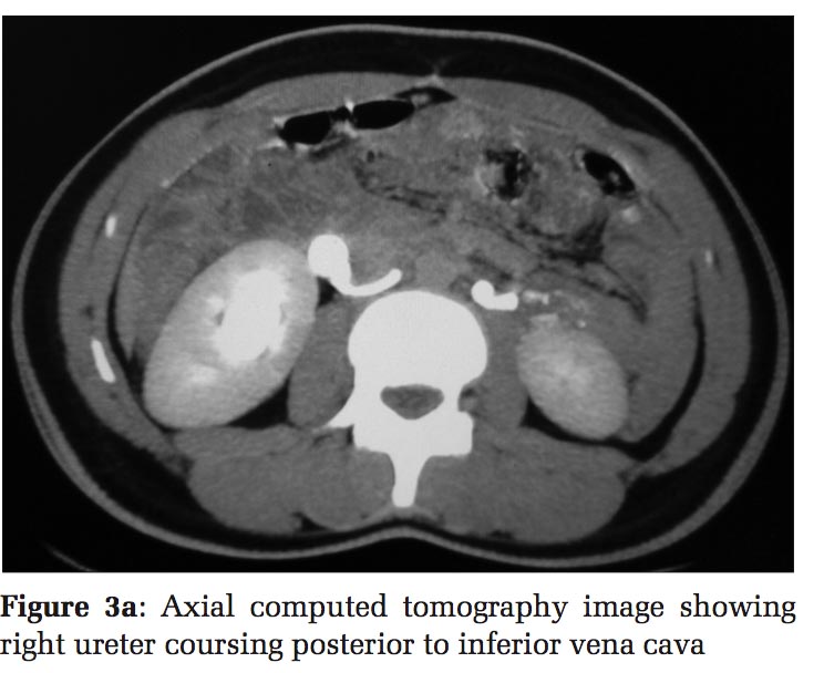
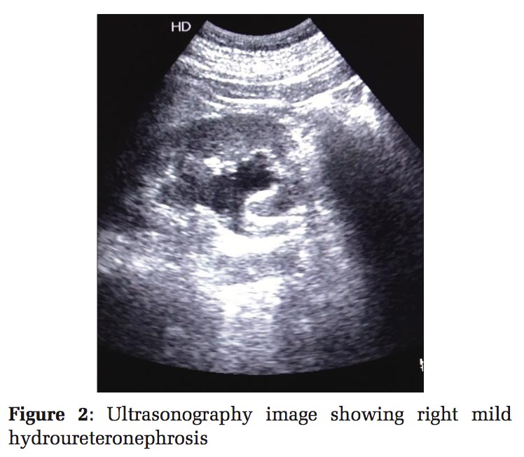
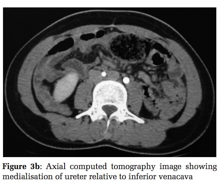
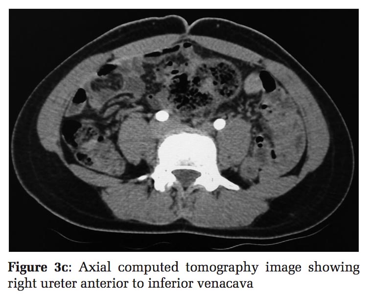
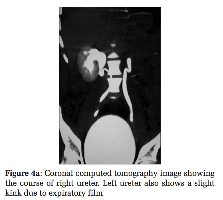
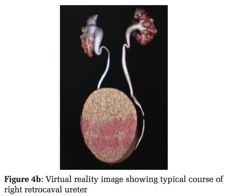
A 20-year-old female patient presented to us with a history of recurrent flank pain for 3–4 years. It was not associated with fever or vomiting. No history of any trauma was there. Her vitals were normal with normal blood counts. She had undergone USG thrice before. One of the reports suggested right hydronephrosis, while the rest two were normal [Figure 1]. We did an US which revealed right hydronephrosis with proximal hydroureter [Figure 2]. No significant abnormality was detected in the rest of the abdomen. Bowel gas obstructed the visualization of entire right ureter. We advised a CT scan to rule out any ureteric calculus. CT scan showed right ureter crossing posterior to inferior vena cava (IVC) [Figure 3a-c]. CECT scan with delayed images confirmed the diagnosis of retrocaval ureter [Figure 4a and b]. Surgery with ureteric repositioning and end-to-end anastomosis was done in the Urology Department, and the patient is doing well on follow-up.
The name retrocaval ureter (also known as circumcaval ureter) is misleading. It sounds like a defect in the ureter, but in reality, the defect lies in the IVC. It is a rare development anomaly of the IVC with a prevalence of 0.9 in 1000 autopsy cases.[1] Here, the ureter passes posterior to the IVC, again to cross anteriorly below.
The IVC develops from three main embryonic veins: Subcardinal vein, supracardinal vein, and the posterior cardinal vein. The subcardinal vein forms the suprarenal part of IVC, while the infrarenal part of IVC is formed by the supracardinal vein. Sometimes the right posterior cardinal vein persists and forms the infrarenal part of IVC. This anomalous persistence of posterior cardinal vein anterior to ureter leads to development of retrocaval ureter. This anomalous persistent part of the subcardinal vein crosses anterior to the ureter, resulting in the development of the retrocaval ureter.[1-3]
In 1983, Hochstetter reported the first case of retrocaval ureter. Nielsen reported an incidence of retrocaval ureters as 0.9 in 1000 in a postmortem series.[2,4,5] In past, the imaging characteristics of retrocaval ureter have been repeatedly described by Abehouse and Tawkin in 1952, Muller and Engel in 1952, Goodwin et al., in 1957, and Rowland et al., in 1960. First, successful surgical correction of this entity was done by Kimbrough.[1,6]
As the IVC develops on the right, the retrocaval ureter is usually present in the right side. However, few cases of the left retrocaval ureter with situs inversus have been reported by Brooks in 1962 and Watanbe et al., in 1991, and its association with left-sided IVC also has been reported Pierro et al. in 1990. Another rare association of bilateral retrocaval ureter with duplicated IVC has been demonstrated by Chou et al., in 2006.[4,7-9]
It is found to be more common in men than women with a ratio of 2.8:1 and usually presents in the 3rd–4th decades. It is mostly asymptomatic, but it can cause obstruction of the ureter, resulting in urinary stasis. This may lead to recurrent episodes of urinary tract infection, flank pain, hematuria, or stone formation.[1-4]
Radiologically, it is classified into two types:
Imaging modalities such as intravenous urography, retrograde pyelography, USG, CT urography, and MRU are useful in the diagnosis and evaluation of the retrocaval ureter. While modalities such as intravenous urography or retrograde pyelography are losing their popularity nowadays, CT and MRU are gradually becoming the investigation of choice for these anomalies.[1,2,10,11]
Intravenous urography demonstrates dilatation of renal pelvis, calyces, and the ureter proximal to the site of obstruction. However, it fails to demonstrate the part of ureter crossing behind the IVC. However, retrograde pyelography can depict the typical fish hook or sickle-shaped deformity of the upper ureter as well as the retrocaval part of the ureter.[10-12]
USG shows hydronephrosis and proximal hydroureter. Abdominal CT is now considered the diagnostic modality of choice for the detection of retrocaval ureter. Being non-invasive, CECT with delayed scan is an appropriate choice of imaging to outline the course of the ureter. Patients allergic to iodinated contrast agents or with poor renal function can undergo MRU with additional advantage of lack of radiation exposure.[10-12]
Asymptomatic patients are usually managed conservatively, while surgical approach with ureteric reposition and end-to-end anastomosis is implicated in symptomatic and complicated cases.[2-4,10,11]
Retroperitoneal fibrosis causing hydroureteronephrosis may mimic retrocaval ureter; however, it is mostly bilateral and CT or MRU can differentiate between the two.[1,10]
Retrocaval ureter is an uncommon cause of unilateral hydroureteronephrosis. CT urography and MRU take the upper hand in diagnosing retrocaval ureter for timely detection and proper management.
To the patient.
Subscribe now for latest articles and news.