Journal of Medical Sciences and Health
DOI: 10.46347/jmsh.v10.i3.24.176
Year: 2024, Volume: 10, Issue: 3, Pages: 275-283
Original Article
Saloni Mahajan1 , Munesh Sangwan2 , Komal Yadav1 , Snehil Agrawal1 , Roopali Sehrawat1 , Rajeev Sen3
1Assistant professor, Department of Pathology, FMHS, SGT University, Gurugram, Haryana, India,
2Medical officer, District Civil Hospital, Charkhi Dadri, Haryana, India,
3Professor and head, Department of Pathology, FMHS, SGT University, Gurugram, Haryana, India
Address for correspondence:
Snehil Agrawal, Assistant professor, Department of Pathology, FMHS, SGT University, Gurugram, Haryana, India.
E-mail: [email protected]
Received Date:04 June 2024, Accepted Date:30 August 2024, Published Date:18 September 2024
Background: The Milan System for Reporting Salivary gland cytopathology is mainly designed for risk stratification. In our study, we categorized cases of fine needle aspiration (FNA) of salivary gland lesions by applying the MSRSGC for the cytological diagnosis and calculating the risk of malignancy (ROM). Methods: Fine-needle aspiration of salivary glands performed for a period of 2 years were recouped. FNA Results were categorized according to the Milan system and correlated with corresponding histopathological follow-up. Risk of malignancy for each diagnostic category was calculated. Results: A total of 54 FNAC of salivary gland lesions were evaluated retrospectively and categorized as: non-diagnostic (ND)-6 (11.11%), non-neoplastic (NN)-12 (22.22%), atypia of undetermined significance (AUS)-1 (1.85%), benign neoplasms (BN)- 18 (33.33%), salivary gland of uncertain malignant potential (SUMP)- 2 (3.70%), suspicious for malignancy (SM)- 1 (1.85%), and malignant (M) 14 (25.92%). Histopathological follow-up was available for 32 of 54 cases (40.4%). Sensitivity, specificity, positive predictive value, negative predictive value and diagnostic accuracy were calculated as 87.50%, 100.00%, 100.00%, and 88.89%, 93.75 respectively. ROM for every category was ND-50%, NN- 0%, AUS- 0%, BN-6.6%, SUMP-100%, SM- 100%, and M-100%. Conclusion: Fine-needle aspiration is a precise diagnostic tool for salivary gland lesions. However, some cases with unusual cytology and overlapping features will have diagnostic and management difficulties.
Keywords
FNAC, Milan system, Risk of malignancy, Salivary gland
Fine-needle aspiration cytology (FNAC) is one of the important and well-accepted techniques for diagnosing salivary gland lesions and it is useful in differentiating non-neoplastic lesions from neoplastic, and benign from malignant neoplasms accurately. 1, 2, 3, 4, 5, 6 Sometimes morphologic features of salivary gland tumours are imbricating and heterogeneous with occasional unusual presentations, a precise diagnosis can be challenging. The American Society of Cytopathology and the International Academy of Cytology proposed an international classification system to conquer this challenge intending to bring similarity in the reporting on FNAC of salivary gland lesions named the Milan System for Reporting Salivary Gland Cytopathology (MSRSGC) in 2018, which was designed to imitate the advantages of The Bethesda System for reporting cervical and thyroid cytology. 7 The MSRSGC is a much-needed universal format for reporting salivary gland cytopathology into reproducible diagnostic categories for a more objective malignancy risk stratification. It enhances the efficacy of salivary gland FNA and allows better communication amongst the cytopathologist and that between the cytopathologist and the treating clinicians.
The second edition of MSRSGC, published in July 2023 includes definition, morphological criteria, and explanations for each of the diagnostic categories and refined risk of malignancy based on the numerous discussions, careful review, and analysis (Table 1). 8
|
Diagnostic category |
Risk of malignancy |
||
|
Category I |
Non-Diagnostic |
15% |
|
|
Category II |
Non-Neoplastic |
11% |
|
|
Category III |
Atypia of Undetermined Significance (AUS) |
30% |
|
|
Category IV |
Neoplasm |
IV A) Benign |
< 3% |
|
IV B) Salivary Gland Neoplasm of Uncertain Malignant Potential (SUMP) |
35 % |
||
|
Category V |
Suspicious for Malignancy |
83% |
|
|
Category VI |
Malignant |
> 98% |
|
It includes new chapters dedicated to application of salivary gland imaging, latest ancillary studies, clinical management, and histological diagnosis including updates in recent WHO 5TH edition of head and neck tumours released in 2022. 9
Present study correlates the findings of fine needle aspiration smears of salivary gland lesions carried out at our institute during a period of 2 years with histopathological diagnosis wherever possible. The FNA diagnosis were categorized in accordance with second edition of MSRSGC, 2023 and histopathological reporting was done in accordance with the WHO 5th edition of head and neck tumours, 2022.
The study was done in the Department of Pathology, SGT Medical College and Research Institute, Gurugram, Haryana. A retrospective study was done where all cases of salivary gland FNAC performed from 1 April 2022 to 31 March 2024 were retrieved from the departmental archives. These salivary gland lesions were earlier aspirated and two to three slides were prepared and stained with Giemsa stain and Papanicolaou stain for routine cytopathology reporting. Fine-needle aspiration cytology diagnosis was retrospectively classified according to the second edition of MSRSGC, 2023 into the following categories: ND - Non-Diagnostic, NN - Non-Neoplastic, AUS-Atypia of undetermined significance, BN - Benign Neoplasm, SUMP - Salivary gland neoplasm of uncertain malignant potential, SM - Suspicious for malignancy and M - Malignant. Along with the cytological diagnosis, age and gender of the patients and site of FNAC were also recovered from the departmental records.
Histopathological diagnosis in all the available cases with surgical excision were also retrieved and the cyto-histopathological correlation was performed. Surgically resected specimens for histopathological examination were received in 32 cases. Slides were stained with hematoxylin and eosin stain and categorization were done according to the WHO classification. Histopathology diagnosis was classified into non-neoplastic, benign and malignant. Both cytology smears and histology slides were re-examined to verify the diagnosis. Results of cytology and histopathology were compared, and the malignancy risk was calculated.
Data was entered in Microsoft Excel, version 2309 and statistical analysis was performed in IBM SPSS Statistics for Windows, version 20.0, NY. The sensitivity, specificity, positive predictive value (PPV) and negative predictive value (NPV) of FNAC were also calculated.
54 cases of fine needle aspiration of the salivary gland were included in the present study, of which 21 were males (38.8%) and the rest 33 were female (61.2%) [Table 2].
|
Gender |
|
|
Male |
21 (38.8%) |
|
Female |
33 (61.2%) |
|
Total |
54 (100%) |
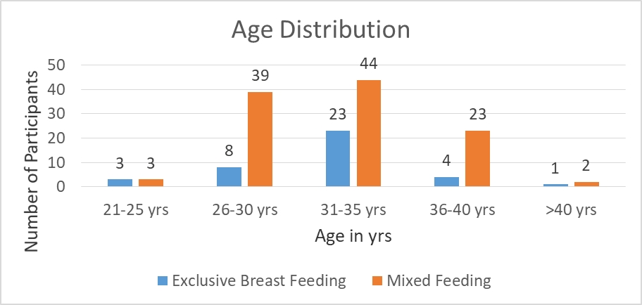
The age range of the patients were from 12 to 78 years with a mean of 46 years. The frequent age group involved was 41 to 50 years with 22 (40.74%) cases followed by 51 to 60 years with 14 (25.9%) cases.
Parotid gland was most commonly involved constituting of 36 (66.66%) cases, followed by the submandibular constituting 12 (22.22%) cases, followed by the minor salivary glands constituting 4 (7.4%) cases, and the sublingual gland comprising of 2 (3.7%) cases [Table 3].
|
Gland Involved |
|
|
Parotid gland |
36 (66.66%) |
|
Submandibular gland |
12 (22.22%) |
|
Sublingual gland |
2 (3.7%) |
|
Minor salivary glands |
4 (7.4%) |
Cytological diagnosis was broadly categorized according to the proposed Milan system for reporting salivary gland cytopathology [Table 4 ] and subcategorized according to the final diagnosis [Table 5].
|
Cytologic diagnostic category |
Total number of cases (n = 54) |
Cases with histopathology follow up (n = 32) |
|
Non-Diagnostic |
6 (11.11%) |
2 |
|
Non-Neoplastic |
12 (22.22%) |
0 |
|
Atypia of undetermined significance |
1 (1.85%) |
1 |
|
Benign Neoplasm |
18 (33.33%) |
15 |
|
Salivary gland neoplasm of uncertain malignant potential. |
2 (3.70%) |
1 |
|
Suspicious for malignancy |
1 (1.85%) |
1 |
|
Malignant |
14 (25.92%) |
12 |
|
Cytologic diagnostic category |
Total number of cases (n = 54) |
Subcategories |
Total number of cases (n=54) |
|
Non-Diagnostic |
6 (11.11%) |
|
|
|
Non-Neoplastic |
12 (22.22%) |
Mucocele |
2 |
|
Chronic sialadenitis |
3 |
||
|
Acute sialadenitis |
1 |
||
|
Sialadenosis |
2 |
||
|
Granulomatous sialadenitis |
1 |
||
|
Reactive lymphadenitis |
3 |
||
|
Atypia of undetermined significance |
1 (1.85%) |
|
|
|
Benign Neoplasm |
18 (33.33%) |
Pleomorphic adenoma |
9 |
|
Warthin's tumor |
4 |
||
|
Basaloid neoplasm |
2 |
||
|
Oncocytoma |
2 |
||
|
Lipoma |
1 |
||
|
Salivary gland neoplasm of uncertain malignant potential |
2 (3.70%) |
|
|
|
Suspicious for malignancy |
1 (1.85%) |
|
|
|
Malignant |
14 (25.92%) |
Mucoepidermoid carcinoma |
8 |
|
Adenoid cystic carcinoma |
2 |
||
|
Salivary duct carcinoma |
2 |
||
|
Acinic cell carcinoma |
1 |
||
|
Non-Hodgkin’s Lymphoma |
1 |
Non-Diagnostic: The adequacy criteria are not well defined for salivary gland FNAC; however, it has been recommended that low cellular smears cause higher discrepancy rates. 4 A total of six cases were categorized under this. In this category, smears had a very scant cell population or necrotic cell debris only. In some cases, only cystic aspirate yielded, and no cytological diagnosis could be furnished.
Non-Neoplastic: This group comprised of twelve cases. The smears did not show any cytological evidence of neoplasm. There were only acinar or ductal epithelial cells with or without an inflammatory background.
Atypia of undetermined significance: This category included one case where the cellular aspirate did not confirm a neoplasm both quantitatively as well as qualitatively which was later diagnosed as non-neoplastic on histopathology.
Benign Neoplasms: Most cases comprising of eighteen cases were placed in this category. Pleomorphic adenoma and Warthin's tumor constituted majority of cases with nine and four cases respectively. Pleomorphic adenoma and Warthin's tumor had clear cytologic and definitive criteria for diagnosis. Other neoplasms appearing benign had a fibrillary or myxoid stroma and were categorized as either basaloid or oncocytic neoplasms with two cases each followed by one case of lipoma.
Salivary gland neoplasm of uncertain malignant potential (SUMP): Smears that were highly cellular (>75%), with a moderate amount of atypia, were grouped under this category. Two cases were included in this category.
Suspicious for malignancy: One case was reported in this category. Cytological findings were suggestive of malignancy but were qualitatively or quantitative insufficient for definitive diagnosis of any carcinoma or malignant neoplasm of the salivary gland.
Malignant: Mucoepidermoid carcinoma (MEC) was the most reported malignancy, eight of fourteen cases, in this study followed by adenoid cystic carcinoma and salivary duct carcinoma with two cases each, followed by one case each of acinic cell carcinoma and Non–Hodgkin lymphoma.
Histopathology diagnosis was received for 32 cases and the spectrum of lesions on histopathological examination was studied [Table 6] [Figure 1, Figure 2, Figure 3, Figure 4, Figure 5]. Correlation was done between cytological and histopathological diagnosis and malignancy risk was calculated for each category. Overall malignancy risk was highest (100%) in SUMP, Suspicious of malignancy and Malignant lesions and lowest (0%) in Non-neoplastic and Atypia of undetermined significance [Table 7].
|
Cytologic category (MILAN system) |
Benign Lesion on Histopathology |
Malignant Lesion on Histopathology |
|
Non-Diagnostic (2) |
Chronic Sialadenitis |
Mucoepidermoid Carcinoma |
|
Non-Neoplastic |
|
|
|
Atypia of undetermined significance (1) |
Chronic Sialadenitis |
|
|
Benign Neoplasm (15) |
Pleomorphic adenoma (7) Warthin's tumor (3) Basaloid neoplasm (1) Oncocytoma (2) Lipoma (1) |
Adenoid Cystic Carcinoma (1) |
|
SUMP (1) |
|
Carcinoma Ex-Pleomorphic Adenoma |
|
Suspicious for malignancy (1) |
|
Salivary duct Carcinoma |
|
Malignant (12) |
|
Mucoepidermoid carcinoma (7) Adenoid cystic carcinoma (2) Salivary duct carcinoma (1) Acinic cell carcinoma (1) Non-Hodgkin’s Lymphoma (1) |
|
Cytologic category
|
Histopathologic category
|
||||
|
Malign ant neoplasm
|
Benign neoplasm
|
Non-neo plastic
|
Total
|
Maligna ncy Risk (%)
|
|
|
Non-Diagnostic |
1 |
0 |
1 |
2 |
50 |
|
Non-Neoplastic |
0 |
0 |
0 |
0 |
0 |
|
Atypia of undetermined significance |
0 |
0 |
1 |
1 |
0 |
|
Benign Neoplasm |
1 |
14 |
0 |
15 |
6.6 |
|
SUMP |
1 |
0 |
0 |
1 |
100 |
|
Suspicious for malignancy |
1 |
0 |
0 |
1 |
100 |
|
Malignant |
12 |
0 |
0 |
12 |
100 |
On cyto-histo correlation, Sensitivity was calculated at 87.5%, specificity at 100%, positive predictive value at 100%, and negative predictive value at 88.89% [Table 8].
|
Statistical parameters |
Percentage % |
|
Sensitivity |
87.5% |
|
Specificity |
100% |
|
Positive predictive value |
100% |
|
Negative predictive value |
88.89% |
|
Diagnostic Accuracy |
93.75% |
FNAC has shown to be remarkable as an early step in the diagnosis and management of salivary gland pathology. 10, 11, 12 Communication between cytopathologists and clinicians can improve the FNA reporting of salivary gland lesions. This promoted the development of a uniform system for reporting salivary gland FNA, i.e., MSRSGC. 8
In the present study, parotid gland was most commonly involved, constituting 36 (66.66%) cases, followed by the submandibular constituting 12 (22.22%) cases, followed by the minor salivary glands constituting 4 (7.4%) cases, and the sublingual gland comprising of 2 (3.7%) cases. A similar pattern of involvement was seen in the study by Jain R et al. in which out of 80 cases, 54 (67.5%) are of parotid gland, 24 (30%) are of submandibular gland and 2 (2.5%) are of minor salivary gland. 8
In the study by Griffith et al., salivary gland lesion FNAC was categorized into commonest morphological categories of benign, neoplasm of uncertain malignant potential, suspicious, and malignant and interpreted risk of malignancy of different categories, which was in accordance to MSRSGC. 8, 11 We also divided cases into similar categories.
In this study, among 54 FNAs, 11.11% were nondiagnostic, 22.22% were nonneoplastic, atypia of undetermined significance 1.85%, 33.33% were benign, 2% were SUMP, 1.85% were suspicious for malignant neoplasm, and 25.92% were malignant. Related findings were also seen in the studies of Rossi et al. and others. 13, 14, 15, 16, 17
Histological diagnosis of all the follow up case was made according to WHO classification of tumors of Head and neck 5th edition. 9
Category I
Out of the six cases categorized as Non-Diagnostic on FNAC, two cases were followed histopathologically, out of which one turned out to be malignant (Mucoepidermoid carcinoma) and the other was chronic sialadenitis, resulting in malignancy risk of 50 % which is higher than the range given by MSRSGC. This represents a selection bias for ROM, which are calculated based only on cases with available histology. The is due to remission of infectious and inflammatory lesion following antibiotics, thus not requiring histopathological follow up. So, the ROM is therefore likely to be higher. The studies by Tochtermann et al 18 , Lubin et al 19 , and Mazzola et al20 shows the similar effect.
In our study, Cystic degeneration of the tumor and aspiration of cyst fluid only with a background of necrotic debris was the main reason for inconclusive reports. This can be prevented by USG-guided FNA from solid areas of the tumor. Another nondiagnostic case which was reported as chronic sialadenitis on histopathology, may be due to the sampling error and can be averted by taking multiple aspirates.
Category II
In the nonneoplastic category of 12 cases of inflammatory and cystic lesions, none of the cases were followed histopathologically, this may be because of remission. Thus, the ROM of this category comes out to be 0%.
Category III
This category involves atypia of undetermined significance. Only one case was placed in this category, which on histopathology follow up came out to be Chronic Siladinitis. The reason behind the cytopathological categorization may be reactive atypia which can be seen in inflammatory lesions. Thus, the risk of malignancy of this category also came out to be 0%.
Category IVa
Out of the 18 cases placed in the benign neoplasm category, on histopathology follow up of 15 cases, one case diagnosed as pleomorphic adenoma on FNAC, histopathologically came out to be adenoid cystic carcinoma giving the risk of malignancy 6.6%. The similarity in morphology between pleomorphic adenoma and adenoid cystic carcinoma like the presence of hyaline globules and myxoid stroma leads to the wrong interpretation as also seen in the study by Klijanienko and Vielh 21 . More wariness and attention to the cellular details should eliminate this problem.
Category IVb, V, VI
Malignancy risk in SUMP, suspicious for malignancy and malignant categories was 100 % each. Case of SUMP turned out to be carcinoma ex pleomorphic adenoma. This may be due to benign component of pleomorphic adenoma seen along with clusters of atypical cells. One case of suspicious of malignancy turned out to be salivary duct carcinoma.
The risk of malignancy of this study was compared to other similar studies. 8, 14, 15, 16, 17, 18, 22 and was found to be variable in various studies. [Table 9] This variability in various study may be due to heterogeneous FNA sampling and tumor types.
|
Study |
I |
II |
III |
IVa |
IVb |
V |
VI |
|
Our Study |
50 |
0 |
0 |
6.6 |
100 |
100 |
100 |
|
Kala et al (2019) 14 |
25 |
5 |
20 |
4.4 |
33.3 |
85.7 |
97.5 |
|
Rolon et al (2020) 15 |
0 |
0 |
75 |
2.2 |
28.6 |
50 |
100 |
|
Jha et al (2021) 16 |
42.86 |
26.67 |
100 |
10.17 |
0 |
71.42 |
100 |
|
Huang et al (2023) 17 |
16.4 |
9.5 |
24.4 |
2.7 |
34.8 |
85.7 |
100 |
|
Tochterm- ann et al (2023) 18 |
26.7 |
5.7 |
34.0 |
1.1 |
21.8 |
92.0 |
99.2 |
|
Bhushan et al (2023) 22 |
33.3 |
2.5 |
0 |
7 |
0 |
66.6 |
100 |
|
Milan |
15% |
11% |
30% |
<3% |
35% |
83% |
98% |
In their study, Jalal et al. reviewed 37 articles from year 2017 to 2020. The total number of salivary gland lesion FNA cases were 16,394, and 8,468 had histological follow-up, resulting in the mean ROM of 16.9% for category I, 10.5% for category II, 39.3% for category III, 2.9% for category IVa, 39.4% for category IVb, 84.2% for category V, and 97.5% for category VI, which corresponded to the ROM range of MSRSGC. 23 Other metanalysis on 10 randomly selected studies by Wang et al. 24 shows similar ROM in the various categories, accounting for 11.4%, 10.9%, 30.5%, 2.8%, 37.7%, 83.8%, and 97.7% in I, II, III, IVa, IVb, V, and VI categories respectively. 25
On correlation of cytopathology with histopathology, FNA reporting of salivary gland lesion using MSRSGC yielded sensitivity of 87.5%, specificity of 100%, positive predictive value of 100%, and negative predictive value of 88.89%. P value was calculated to be <0.001. These findings are in accordance with various studies done on cytohistological correlation. Many publications also have chronicled the sensitivity, and specificity of 87%–100%, which might be caused by the heterogeneous FNA sampling and due to morphologically similar tumor types and observer experience [Table 10].14, 15, 16, 23, 26, 27
|
Study |
Sensitivity |
Specificity |
Positive predictive value |
Negative predictive value |
Diagnostic Accuracy |
|
Our study |
87.5% |
100% |
100% |
88.89% |
93.75% |
|
Jain et al. (2013) 23 |
92.8% |
93.9% |
81.2% |
98.4% |
---- |
|
Rohilla et al (2017) 26 |
79.4% |
98.3% |
96.4% |
89.2% |
91.4% |
|
Kala et al. (2019) 14 |
83.33% |
98.31% |
95.74% |
92.80% |
93.60% |
|
Rolon et al. (2020) 15 |
93.3% |
94.6% |
82.4% |
98.2% |
94.4% |
|
Jha et al. (2021) 16 |
64.28% |
97.01% |
90% |
86.67% |
87.37% |
|
Hindi et al. (2022) 27 |
78.3 |
98.0% |
94.7 |
90.0 |
---- |


Fine needle aspiration cytology is a steady, safe, reliable, economically practical technique in diagnosing the salivary gland lesions. However, globally diagnostic complexity and the heterogeneous approach to the salivary gland tumors needs a risk-based broad stratification categorization for productive managements. The Milan classification tries to address this requirement by providing an effective six-tiered initial grading system while providing the cytopathologist with an easy template-based classification system, which marginalizes diagnostic discrepancies to a minimum.
Subscribe now for latest articles and news.