Journal of Medical Sciences and Health
DOI: 10.46347/jmsh.v9i1.22.317
Year: 2023, Volume: 9, Issue: 1, Pages: 57-63
Original Article
S Megha1 , Indradhanush 2 , D Venkatesha3
1Assistant Professor, Department of Microbiology, AIMS, Karnataka, India,
2Undergraduate student, AIMS, Karnataka, India,
3Prof and Head,Department of Microbiology, AIMS, Karnataka, India
Address for correspondence:
S Megha, Assistant Professor, Department of Microbiology, AIMS, Karnataka, India.
E-mail: [email protected]
Received Date:01 September 2022, Accepted Date:30 January 2023, Published Date:08 March 2023
Background: Plants have been the basis for many medicinal compounds that are used in modern-day medicine, screening for medicinal plants for bioactive compounds is a way of identifying and developing new antimicrobials that are less expensive and with improved safety and efficacy. Objective: The objective of this study was to investigate the in vitro antimicrobial activity of Azadirachta indica (neem) using leaf extracts of Ethanol, Petroleum ether, and Ethyl acetate and testing them on E.coli, Klebsiella, S.aureus, MRSA and C.albicans. Materials & Methods: Minimum inhibitory concentration (MIC) was found using the serial dilution method and antimicrobial susceptibility testing (AST) was done using the disc diffusion method. Results: Ethanolic extract showed better results against Klebsiella and S.aureus, Ethyl acetate extract showed better results against E.coli and MRSA, whereas both ethanolic and petroleum ether extract showed activity against C.albicans. 2,2-diphenyl-1-picryl-hydrazyl-hydrate (DPPH) assay showed that all the extracts have antioxidant property and variations were noted in the antimicrobial activity on comparing samples taken from different locations. Conclusion: Further studies are needed into the bioactive compounds of neem in order to explore their antimicrobial activity.
Keywords: Antimicrobial susceptibility, Azadirachta indica, Ethanol, Ethyl acetate, Disc diffusion, MIC, Petroleum ether
Use of herbal medicines in the developed world continue to rise because they are rich source of novel drugs and their bioactive principles form the basis in medicine, 1 with few adverse reactions. The emergence and dissemination of multidrug resistance bacteria has made chemically synthesized antibiotics ineffective. 2 These circumstances have propelled researchers and scientists to explore new antimicrobial substances from various sources such as medicinal plants. 3
Azadirachta indica is an evergreen tree commonly found in the Indian subcontinent, which is famous by the local name ‘Neem’, it has been considered to provide freedom from all diseases, and used for thousands of years in Indian and African continents. Different parts of the plant including flowers, leaves, seeds and bark have been used to treat both acute and chronic human diseases; and used as an insecticide; antimicrobial, larvicidal, antimalarial, antibacterial, antiviral, and spermicidal 4. Neem tree contains large number of biologically active compounds that are diverse and complex. More than 140 compounds including alkaloids, flavonoids, triterpenoids, carotenoids and nimbin have been isolated from different parts of the plant 5, 6 . The extracts of the tree (leaves, roots, bark) have been reported to possess anti-viral, anti-fungal, anti-bacterial properties7. The purpose of this study is to investigate the antimicrobial property of neem using solvent extracts method on neem leaves.
To obtain extract of Neem leaves using Ethanol, Petroleum ether (Pet.ether) and Ethyl acetate from two geographical area.
To investigate the antimicrobial activities of the above-mentioned extracts of Neem leaves against ATCC organisms, Escherichia coli (ATCC 25922), Staphylococcus aureus (ATCC 25923), Klebsiella pneumoniae (ATCC 13883), Methicillin-resistant Staphylococcus aureus (MRSA ATCC 33591) and clinical isolate of Candida albicans
Determination of MIC and comparison of various solvent extracts of neem leaves for its antimicrobial property against above-mentioned organisms.
The present study is an observational study conducted in the department of Microbiology at AIMS, B.G.Nagara for a period of two months.
Test organisms used are Escherichia coli (ATCC 25922), Staphylococcus aureus (ATCC 25923), Klebsiella pneumoniae (ATCC 13883) and Methicillin-resistant Staphylococcus aureus (MRSA ATCC 33591), obtained by retrieving the ATCC stock culture suspensions maintained in the Microbiology department. Candida albicans (C.albicans)was taken from patient sample collected in Adichunchanagiri institute of medical sciences, B G Nagara as ATCC strain was not available at the period of conduction of study.
Fresh leaves of Azadirachta indica were collected from the (1) vicinity of Adichunchanagiri university area and from (2) Mysore university area. Both the samples of leaves were washed gently with tap water and left to dry at room temperature for 5 days, then University sample was labelled as ‘ACU Neem’ and that collected from Mysore University as ‘UOM Neem’.About 10g of the powdered sample is weighed in a 50ml graduated tube and corresponding solvent is filled till 50ml mark.
|
Solvent / Sample |
Petroleum ether |
Ethanol |
Ethyl acetate |
|---|---|---|---|
|
ACU Neem (weight in gm) |
9.9596 |
9.9924 |
9.9407 |
|
UOM Neem (weight in gm) |
9.9681 |
9.9865 |
9.9746 |
Then the solvent containing neem powder is fixed onto the rotator and mixed for about 1 day, after which the contents of each tube were filtered using Whatman no.1 filter paper. The residue is subjected to the same solvent extraction again by adding solvent to it then the process is repeated for all the samples. The obtained filtrate is then transferred onto a glass petri dish and left to dry out so that the solvent evaporates leaving behind the fine extracts 8.

The dry powder extracts of Azadirachta indica obtained from the above process is scrapped off from the plates and then dissolved in dimethyl sulfoxide (DMSO).
The minimum inhibitory concentration of a compound is defined as the lowest concentration of that compound required to inhibit the growth of the microorganism. All bacterial strains are inoculated to Muller Hinton broth and fungal culture into Sabouraud’s dextrose broth under sterile conditions and then incubated in the incubator at 35-37ºC for 24hrs after which the optical density (OD) is measured using an electro spectrophotometer to obtain OD of 4-6 if OD is low incubatory period is extended 1 .
The final concentration of the stock solution of extracts after dissolving in DMSO is 1.024µg/100µl. Sterile Microtiter plates are used for MIC dilution,25µl of broth is added to 2-8,11th (media control) and 12th (growth control) wells, then 25µl of drug is added to 1st (drug control 256µg) and 2nd well. Drug dilution is carried out from 2nd (128µg) to 9th (2µg) well. 25µl of solvent is added to 10th well (solvent control) then to each well 25µl of suspension of the organism is added from 1st to 9th well and 12th well (growth control), 25µl of un-inoculated broth is added to 11th well (media control). Then the microtiter plates are incubated in incubator at 37ºC for 24hrs after which OD is measured.
The results are then read, the well with the lowest drug concentration showing maximum inhibition was recorded, and this concentration of the extracts is referred to minimum inhibitory concentration (MIC).
After MIC to confirm whether the extract is bacteriostatic or Bactericidal MBC was done,
5µl of contents in the next consecutive well to its left with more concentration along with MIC well and 12th well are inoculated onto MHA plates and incubated 18-24hrs to see for growth of the organisms. The concentration of the well before the MIC well with more concentration showing no growth is recorded as MBC value 9.
It is used to predict antioxidant activities by mechanism in which antioxidants act to inhibit lipid oxidation, (scavenging of DPPH radical) and therefore determine free radical scavenging capacity. DPPH is violet and reduced in the Presence of antioxidant molecule to give rise to colorless ethanol 10.
Preparation of sample drug - Add 100µl sample drug to 900µl DMSO
Preparation of reagent - DPPH 2mg; 100% Ethanol-25ml
Preparation of tris buffer - 0.25ml of water.
The whole procedure is carried out in a dark place.
Keep incubation for 30min at room temperature in dark place, then check OD at 517nm. 11
The antibacterial activity of the solvent extracts of Azadirachta indica is done using agar disc diffusion method. In this method (Kirby Bauer) the impregnated discs diffuse the antibiotics into the surrounding agar medium. As the distance from the disc increases the concentration of the antibiotic decreases 12.
3.5g of Muller Hinton agar is added to 100ml of distilled water and autoclaved at 121ºC for 15 minutes at 15lbs and poured in sterile petri plates of 90mm diameter up to uniform thickness and the agar is allowed to set at room temperature.
All bacterial strains are inoculated to Muller Hinton broth and fungal culture into Sabouraud’s dextrose broth under sterile condition then incubated in the incubator at 35-37ºC for 24hrs after which the optical density (OD) is measured using an electro spectrophotometer to obtain OD of 4-6 if OD is low incubatory period is extended.
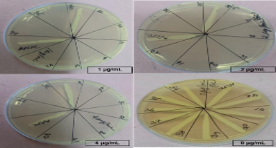
Once the desired OD is obtained the culture suspensions were inoculated on to sterile MHA plates prepared earlier using lawn culture method using a sterile cotton Q-tip. The cotton is inserted into the suspension and rotated and compressed against the wall of the test tube to express the excess fluid. To ensure growth is uniform and confluent the cotton tip is streaked in zig-gag fashion then turning the plate by 60º repeating the same 3-4 times to leave no gap. The discs are prepared using Whatman filter paper measuring 6mm in diameter and there sterilized by hot air oven. The discs are then placed at appropriate position with the center disc being the standard antibiotic. With the observation and values obtained from the above MIC experiment suitable six consecutive dilution are taken, such that it includes the MIC concentration. The six chosen concentrations are loaded on to the discs in order of their dilution using micropipette. An extra disc labelled ‘C’ is used as control which is loaded with DMSO and at the center standard disc of Gentamicin GM10 is used as positive control, all the above steps are done under sterile condition to avoid contamination and same is done for each of the chosen organisms. Inoculated MHA plates are then placed inside incubator at 35-37ºC for 24hrs. The results are then read by measuring the zone of inhibition for each of the discs using a scale and the obtained data is tabulated.
Data collected was entered in excel sheet and analysed using SPSS software, results were expressed in terms of descriptive statistics.
|
Extract |
E.coli |
Klebsiella |
S.aureus |
MRSA |
C.albicans |
|
ACU Neem Ethanol |
16 |
32 |
32 |
32 |
64 |
|
UOM Neem Ethanol |
32 |
32 |
32 |
32 |
64 |
|
ACU Neem Petroleum Ether |
32 |
32 |
32 |
64 |
64 |
|
UOM Neem Petroleum ether |
64 |
32 |
64 |
32 |
64 |
|
ACU Neem Ethyl Acetate |
32 |
32 |
64 |
16 |
64 |
|
UOM Neem Ethyl Acetate |
64 |
32 |
16 |
32 |
64 |


|
EXTRACT |
E.coli |
Klebsiella |
S.aureus |
MRSA |
C.albicans |
|
ACU Neem Ethanol |
11 |
12 |
6 |
6 |
6 |
|
UOM Neem Ethanol |
11 |
16 |
8 |
10 |
7 |
|
ACU Neem Petroleum ether |
13 |
13 |
6 |
8 |
6 |
|
UOM Neem Petroleum ether |
12 |
14 |
0 |
13 |
7 |
|
ACU Neem Ethyl Acetate |
14 |
12 |
0 |
10 |
0 |
|
UOM Neem Ethyl Acetate |
12 |
13 |
4 |
13 |
6 |

In recent years, we have seen antimicrobial resistance rapidly emerge at a global scale and spread from one country to the other faster than previously thought. Multidrug-resistant bacteria are endemic in many parts of the world. There is no question that the widespread use, overuse, and misuse of antimicrobials during the previous years have been associated with the explosion of antimicrobial resistance 11 . Use of herbal medicines in the developed world continue to rise because they are rich source of novel drugs and their bioactive principles form the basis in medicine 1. These circumstances have propelled the researchers and scientists to explore new antimicrobial substances from various sources such as medicinal plants. 3
In the present study, leaves of Azadirachta indica also known as ‘Neem’ was taken to test their potential as antimicrobial agent. Solvent extraction was done using three solvents namely Ethanol, Ethyl acetate and Petroleum ether. These solvent extracts were tested against two gram- negative (Escherichia coli and Klebsiella) two gram-positive organisms (Staphylococcus aureus and MRSA) and a fungus (Candida albicans).
The results obtained from minimum inhibitory concentration testing (Table 2) showed that Ethanolic extract of the leaves had more activity against E. coli (16 and 32µg) than the other two solvent extracts, however all the extracts showed equal activity against Klebsiella (32µg). For S.aureus ethyl acetate extract gave the best result (16µg) followed by ethanolic extract (32µg). Whereas for MRSA best results were seen with Ethyl acetate (16 and 32µg) followed by ethanolic extract (32µg). For Candida albicans all showed similar results (64µg) (Figure 2). In a study conducted by Rajaratna Reddy et al, showed that aqueous extracts also have good antimicrobial property After MIC to confirm whether the extract is bacteriostatic or bactericidal MBC was done , then it was confirmed that the extracts had bactericidal action. DPPH assay showed that extracts have antioxidant property and UOM Neem Pet.ether has the highest antioxidant property when compared to other extracts.
Taking MIC values as reference antimicrobial susceptibility testing (AST) was done by disc diffusion method. Alternatively in a study conducted by Mahmood Rasool et al., they used well diffusion method and had achieved good results. The zone of inhibition was measured (Table 3) after incubation the results showed Ethyl acetate extracts showed best result among the three solvents (ZOI 14 and 12mm) followed by Petroleum ether (12 and 13mm) for E. coli, however maximum zone was seen with ethanolic extract for Klebsiella (16mm) followed by Petroleum ether and Ethyl acetate. For S.aureus good results were seen with ethanolic extracts (8 and 6mm) followed by Ethyl acetate (7and 0mm) but Petroleum ether failed to inhibit the organism(0mm). Whereas for MRSA good results were seen with Ethyl acetate and petroleum ether (13&10mm and 13& 8mm) followed by the ethanolic extract (10 and 6mm). For Candida albicans ethanol and Petroleum ether showed better results (6 and 7mm) than ethyl acetate (Figure 4).
From the above data on comparing both sample ‘ACU Neem’ and sample ‘UOM Neem', Sample ACU neem showed better antimicrobial activity against E. coli, UOM Neem showed better antimicrobial activity against Klebsiella spp. For S.aureus both showed almost similar results, but for MRSA sample UOM neem showed slightly better results than ACU Neem. For Candida albicans all extracts showed almost similar results. From this it is evident that site of sample collection also has significant impact on the antimicrobial activity of the extracts. The altitude, cultivar fertility, soil conditions and climatic conditions from where plant sample is collected all have impact on the antimicrobial activity. 13
Ethanolic extract showed better results against Klebsiella spp and S.aureus, Ethyl acetate extract showed better results against E.coli and MRSA, whereas both ethanolic and Petroleum ether extract showed activity against C.albicans. 2,2-diphenyl-1-picryl-hydrazyl-hydrate (DPPH) assay showed that all the extracts have antioxidant property and variations were noted in the antimicrobial activity on comparing samples taken from different locations. The antimicrobial property of extracts of different solvents showed variations in their antimicrobial activity against different organisms.
The results obtained from minimum inhibitory concentration testing (Table 2) showed that Ethanolic extract of the leaves had more activity against E. coli (16 and 32µg) than the other two solvent extracts, however all the extracts showed equal activity against Klebsiella spp (32µg). For S.aureus ethyl acetate extract gave the best result (16µg) followed by ethanolic extract (32µg). Whereas for MRSA best results were seen with Ethyl acetate (16 and 32µg) followed by ethanolic extract (32µg). Hence MIC of the leaf extracts depends on the solvent used for extraction and also showed varied antimicrobial property against different organisms.
Along with the above-mentioned extracts other solvent extracts like methanol, aqueous extracts can also be tried and has shown significant results in the past experiments. More microbes can also be included to widen the array and obtain more data. Other factors like temperature and pH changes can be included to find the range where the antimicrobial activity is optimum. Since the extracts were crude and methodological choices were constrained by need for higher technology, the actual results gone using better technology is expected to have better and specific antimicrobial activity. To make things more sophisticated further studies can be conducted to isolate different components of the neem extract and test their actions and mechanisms at molecular level on a higher scale.
The limitation of the present study is that since MDR strains were not used in the present study activity against drug resistance cannot be concluded. Further to improvise the study multiple samples can be tested in future with the same solvent extracts to obtain repeatability. Studies are needed into the bioactive compounds of neem in order to explore its antimicrobial activity. Hence Azadirachta indica can be used treatment of commonly encountered organsims in the hospital because of its antimicrobial and anti-oxidant property. Further studies are required to confirm its antimicrobial properties against multidrug resistant organisms. 14
I acknowledge Dr. M G Shivaramu, principal and Dr. Venkatesha D Head of Dpt of Microbiology, Adichunchanagiri Institute of Medical Sciences, B G Nagara. I express my sincere thanks to Dr. Sudhanva KV whose support and technical guideline helped the present work as its show. Thanks Adichunchanagiri Institute for Molecular Medicine (AIMM) for providing material and equipments
Subscribe now for latest articles and news.