Journal of Medical Sciences and Health
DOI: 10.46347/jmsh.v9i3.23.250
Year: 2023, Volume: 9, Issue: 3, Pages: 256-262
Original Article
Vignesh Mahendiran1 , S S Vijay Kumar2 , P Bindhya Ponnuchamy3
1Department of Orthopedics, KS Hegde Medical Academy, Deralakatte, Mangalore, 575018, India
2Department of Emergency Medicine, KS Hegde Medical Academy, Deralakatte, Mangalore, 575018, India
3Department of Anaesthesia, KS Hegde Medical Academy, Deralakatte, Mangalore, 575018, India
Address for correspondence:
S S Vijay Kumar, Department of Emergency Medicine, KS Hegde Medical Academy, Deralakatte, Mangalore, 575018, India.
Email: [email protected]
Received Date:22 July 2023, Accepted Date:02 November 2023, Published Date:19 December 2023
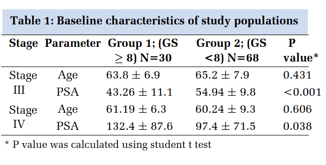
Introduction: High-speed accidents and trauma are becoming increasingly common and often result in forearm fractures in developing countries and even rural populations are not immune to such injuries. To address this issue, there has been a growing trend towards using flexible intramedullary nailing systems, such as the Titanium Elastic Nailing System (TENS). Methodology: This prospective study conducted at PES Institute of Medical Sciences and Research, Kuppam India, between June 2019 and March 2023 after receiving institutional ethical approval, patients with diaphyseal fractures of the forearm were treated with TENS, after their consent and, then followed up at 1st, 3rd, and 6th months post-operatively using Daruwalla's Clinical criteria. Result: The study involved 300 patients, with a male predominance (60%) and most common age group of 11-20 years old (26.7%) involving right forearm (63.3%), at middle third (76.7%) with transverse (80%) and closed (86.7%) nature. The leading cause was road traffic accidents (50%). At the first month, 86.7% had fair outcomes according to Daruwalla's grading, with 40% showing excellent outcomes by the 6th month. Over time, the proportion of patients with good (46.7%) and excellent (40%) outcomes increased, while fair and poor outcomes decreased (13.3%, 0%) Conclusion: TENS has shown efficacy in treating forearm fractures. Positive trends in clinical grading were seen, with a majority of patients achieving fair to excellent outcomes by the 6th month post-operative follow-up. These results support TENS, even in rural settings with limited resources, as a viable alternative to plate osteosynthesis.
Keywords: Intramedullary Nailing, Forearm Injury, Fracture
Treatment of forearm fractures varies based on patient age, fracture pattern, and type of injury (e.g. open or closed). Pediatric fractures are typically managed with non-surgical methods such as slabs and casts, as the growth potential of immature bones enhances union. In contrast, adolescents and adults often require surgical intervention 1, 2.
There is a recent trend towards prompt surgical intervention for diaphyseal fractures, regardless of age and injury pattern. The most common surgical options are plate osteosynthesis and intramedullary nailing. These procedures involve using dynamic compression plates (DCPs) or locking compression plates (LCPs) to achieve good anatomical repair and radial bow restoration. However, these techniques have some drawbacks, such as a longer incision, disruption of periosteal blood vessels, stripping of soft tissues, and interruption of hemostasis at the fracture site, resulting in a higher risk of delayed union 3.
Therefore, there has been a shift towards using advanced surgical methods, such as intramedullary nailing with the Titanium Elastic Nailing System
This study aimed to examine the demographic profile, pattern, and causes of diaphyseal forearm fractures in patients, and evaluate the effectiveness of elastic stable intramedullary nailing using Daruwalla's Clinical Grading, which was performed periodically in rural southern India 4.
This prospective study was conducted on patients presenting to Emergency Room at PES Institute of Medical Sciences and Research, Kuppam India after obtaining ethical clearance from Institutional Review Committee. This study was conducted between June 2019 and March 2023 on patients who sought treatment for displaced diaphyseal fractures of forearm. Individuals with congenital bone disorders or pathological fractures were excluded from the study. The sample size was calculated using the following formula-
n = Z2 x p x q / e2
= 1.962 x 0.5 x 0.5 / 0.062
= 267
Where,
N = minimum required sample size
Z = 1.96 at 95% Confidence Interval (CI)
P = prevalence taken as 50% for maximum sample size calculation
Q = 1-p
E = margin of error, 6%
The required sample size was 265, but we took 300 patients. Once patients were stabilized in Emergency room with principles of advanced trauma life support and major life-threatening injuries were ruled out, X rays of the injured forearm was obtained and the fractures were immobilized with slabs. The patients were explained in detail the pros and cons of the intervention, written consent was taken from all patients who were recruited for surgical procedure. The surgical reduction of the fracture was considered successful if it had a minimum of 50% cortical contact and an angulation of no more than 10 degrees in either the anteroposterior or mediolateral plane during the operation. To immobilize the operated limb, an above-elbow slab was used for two weeks. Clinical evaluations were performed on the patients at 1st, 3rd, and 6th months post-surgery to assess range of motion (using a goniometer) and limb length discrepancy (measured from the tip of the distal radial styloid to the lateral epicondyle), and angular deformities (via radiographs of the forearm). Then the patients were graded according to Daruwalla's Clinical criteria 4
|
Classification |
Criteria of Limitation |
|
Excellent |
Movements equal on both sides |
|
Good |
< 20o of limited rotation on injured side |
|
Fair |
20o-40o of limited rotation on injured side |
|
Poor |
> 40o of limited rotation on injured side |
Inclusion criteria
All patients with diaphyseal fractures of the forearm who underwent flexible intramedullary nailing
Age between 5 years and 60 years
Exclusion criteria
Congenital disorders.
Population with pathological fractures.
Less than 5 years and more than 60 years
Statistical analysis was done by SPSS v 16 statistical software (SPSS Inc., IBM company, New York, US), and the descriptive statistic was used and results were expressed in frequencies and percentages.
The surgical stabilization of the radius and ulna requires separate entry points, one at each end of the forearm. The radial entry point is distal and the ulnar entry point is proximal, guided by an intraoperative image intensifier. To access the radial entry point, a 2-3 cm transverse or longitudinal incision is made over the palpable dorsal tubercle and an awl instrument is used to create an entry, starting perpendicularly and then angling obliquely towards the elbow. To access the ulnar entry point, a 1.5 to 2 cm transverse incision is made over the proximal lateral aspect of the olecranon, 3 cm distal to the apophysis. After reduction is achieved through external manipulation, the appropriate size TENS nail is passed and fixed to the injured bone's metaphysis. The distal end of the nail is cut 5-10mm from the bone before closing the entry point. If the injury is open, the procedure mentioned above is followed. In case of a closed injury with instability, a percutaneous intramedullary nailing is performed without opening the fracture site.
The study involved 300 patients with forearm diaphyseal fractures. The most common age group was 11-20 years old, making up 26.7% of the participants. There was a male predominance, with 60% of the patients being male. 260 patients had sustained fractures in both bones, while 30 had isolated radius fractures and 10 had an isolated ulnar fracture (Figure 1, Figure 2)


The study found that the right forearm was the most frequently affected with 63.3% of patients. The middle third of the forearm was the most commonly affected area, accounting for 76.7% of cases. Most of the fractures were transverse in nature (80%) and closed (86.7%). In the study, road traffic accidents (RTA) were the leading cause of injuries accounting for 50% of cases, while falls from height contributed to 30% and assaults accounted for 20%. Closed reduction was commonly performed in 83.3% and 2.5mm TENS nail was frequently used (50%).
|
Variables |
n |
n% |
|
Side |
||
|
Right |
190 |
63.3 |
|
Left |
110 |
36.7 |
|
Site |
||
|
Distal 3rd |
30 |
10 |
|
Middle 3rd |
230 |
76.7 |
|
Proximal 3rd |
40 |
13.3 |
|
Pattern |
||
|
Oblique |
60 |
20 |
|
Transverse |
240 |
80 |
|
Type |
||
|
Closed |
260 |
86.7 |
|
Opened |
40 |
13.3 |
|
Etiology of Injury |
||
|
Assault |
60 |
20 |
|
Fall From Height |
90 |
30 |
|
Road Traffic Accident |
150 |
50 |
|
Type of Reduction Done |
||
|
Closed Reduction |
250 |
83.3 |
|
Open Reduction |
50 |
16.7 |
|
Diameter of Nail Size Used |
||
|
2.0mm |
30 |
10 |
|
2.5mm |
150 |
50 |
|
3.0mm |
120 |
40 |
At the first month postoperatively, the majority of patients (86.7%) had a fair functional outcome according to Daruwalla's Clinical grading. No patients showed excellent or good outcomes at this time. Over the following months, the proportion of patients with good and fair outcomes shifted, with more patients having a good outcome (86.7% at the 3rd month and 46.7% at the 6th month) and fewer patients having a fair outcome (13.3% at the 3rd and 6th months). By the 6th month, 40% of patients had an excellent functional outcome.
|
Grade |
1 st month |
3 rd month |
6 th month |
|||
|
n |
n% |
n |
n% |
n |
n% |
|
|
Excellent |
0 |
0 |
0 |
0 |
120 |
40 |
|
Good |
0 |
0 |
260 |
86.7 |
140 |
46.7 |
|
Fair |
260 |
86.7 |
40 |
13.3 |
40 |
13.3 |
|
Poor |
40 |
13.3 |
0 |
0 |
0 |
0 |
Forearm fractures are a common type of injury that can cause significant suffering and healthcare costs. According to recent studies, the incidence of forearm fractures is on the rise in developing countries like India 5. Our study found that the majority of forearm fractures occurred in the adolescent age group and in males, which is consistent with previous studies 6. This increase is likely due to a growing number of two-wheelers, particularly among the adolescent population, and a rise in reckless driving The most common pattern of fracture observed in our study was transverse in nature and closed in type, a finding that is supported by previous research 7. The leading cause of forearm fractures in our study was road traffic accidents, followed by falls, and these results are in line with previous studies. When focusing specifically on pediatric age groups, previous studies have found that falls and sports injuries are the leading causes of forearm fractures 8.
Forearm fracture management involves closed reduction and long arm casting in children with fast bone-healing and potential for growth-assisted remodeling, while surgical intervention such as plating or nailing is required in adults as closed methods cannot ensure anatomic reduction and healing 9, 10, 11. The AO group's (ArbeitsgemeinschaftfürOsteosynthesefragen) principles which are: Restoration of anatomy, Stable fracture fixation, Preservation of blood supply, and early mobilization are also applied for management of forearm fracture too 12.
Open reduction and compression plate fixation has been the traditional approach for the management of fractures of the forearm. This method has demonstrated high rates of union and improved range of movement, particularly rotational movement of the forearm 11. However, it also has its own set of limitations, such as a longer incision, disruption of the periosteal blood supply, soft tissue stripping, and disruption of hemostasis at the fracture ends, which can result in a higher incidence of delayed union. In addition, plate fixation also poses the risk of refracture after plate removal, a unique complication. These considerations underscore the importance of exploring alternative treatment options that may address the limitations associated with open reduction and compression plate fixation in forearm fractures.
Thus, intramedullary nailing options like TENS has become a favored approach due to its advantages, including rapid union, low incidence of mal-union, preservation of the growth plate, shorter surgical time, minimal soft tissue disruption, early mobilization, good to excellent range of motion, ease of implant removal, and improved cosmetic outcomes, leading to increased patient satisfaction. Unlike plates, which are fixed externally to the bone, TENS functions as a load-sharing device located closer to the mechanical axis of the bone and has reduced bending loads, resulting in a lower risk of refracture 13. Our study showed no cases of non-union or refracture. However, three patients experienced superficial pin site infections at the ulnar entry site, which were treated with oral antibiotics and did not impact the patients' functional outcomes.
The early studies of intramedullary nailing for the treatment of forearm fractures conducted in France showed promising results 14. The indications for surgery included unstable fractures (26%), failure of conservative treatment (18%), refracture (12%), and initial operative treatment for adolescents (42%). The average age at the time of surgery was 10 years, and curved stainless steel pins with a diameter of 1-3 mm were used. One-year post-operative follow-up results indicated that 98% of patients had a range of motion with no loss greater than 20° compared to the unaffected side, demonstrating the efficacy of intramedullary nailing in the treatment of forearm fractures 14, 15, 16.
The calculation of the necessary nail diameter in intramedullary nailing was determined using Flynn et al's formula, which is based on the width of the narrowest point of the medullary canal as seen on the AP and lateral views. This formula calculates the diameter as 0.4 times the width of the medullary canal 17. This study found that a 2.5mm diameter nail was used in the majority of patients.Patients were evaluated postoperatively using Daruwalla's method to categorize the range of movement at 1st, 3rd and 6th months after TENS nailing. At the first month, none of the patients showed excellent or good outcomes, with 86.7% having a fair outcome. This can be attributed to the fact that the inflammatory and reparative phase of fracture healing typically lasts for 3-4 weeks, making it unlikely for patients to attain good or excellent outcomes during this time 18. However, when the patients returned for their second follow-up at the 3rd month, the majority (86.7%) showed a good outcome, with only 13.3% having a fair outcome. This shift can be attributed to the start of the remodeling phase, the final stage of fracture healing, which usually begins after 6-8 weeks. By the time of their last visit at the 6th month, 46.7% of the patients had a good outcome, while 40% had an excellent outcome, indicating a shift towards better outcomes compared to previous follow-ups.
Studies on the use of TENS for forearm fractures in the pediatric population by Jubel et al, Flynn et al, and Luhmann et al have reported excellent or good result, which used Price's criteria and Anderson criteria for clinical evaluation, which differs slightly from Daruwalla's grading 19, 20, 21. Lascombes and Metaizeau found 92% of their 80 patients had excellent results following intramedullary forearm nailing in both bones 14
Prakriti et al conducted a study among the adult Indian population and found 46.67% of their patients had good outcomes and 30% had excellent outcomes using the Grace and Eversman scoring system 22. Ying-Cheng Huang et al, among the elder Taiwanese population, also concluded that TENS led to better outcomes using the Quick-Dash score, with a score of 7.92 (range 4.5-25, with lower scores indicating better outcomes) 23.
In our study we achieved less than 10 degree postoperatively angulation in all but two patients
The two exceptions were a patient whose nail size was too thin to maintain reduction, but this did not affect their forearm range of movement, and another patient with a proximal third both bone forearm fracture where it was challenging to control the proximal fragments, but again this did not impact their forearm movements. Comparative studies, such as Carmichael's study of 15 patients who underwent internal stabilization of unstable both bones forearm fracture with flexible intramedullary nails versus 16 patients who underwent open reduction and plate osteosynthesis, reported excellent to good results in both groups with only minor complications in the intramedullary group 24. Shah et al also found that the intramedullary group had fewer complications, including none major ones such as nerve injury, compared to the plate osteosynthesis group 25.
Based on the successful outcomes observed in the treatment of unstable and/or open diaphyseal fractures of the forearm bones, we suggest the use of flexible intramedullary nailing as a promising approach for internal stabilization of such fractures.
In conclusion, the study showed that TENS is a successful option for fractures of the forearm. The study population was dominated by males, with the most common age group being 11-20 years old. RTA was the main cause of injury, and most fractures were transverse and closed. The results from the clinical grading indicated a positive improvement in functional outcomes over time, with 86.7% of patients having a fair outcome at the first month post-injury, increasing to 86.7% good outcome at the 3rd month, and 46.7% at the 6th month. By the 6th month, 40% of patients had an excellent functional outcome.
The benefits of using TENS for forearm fractures include its minimally invasive technique, preservation of fracture hematoma, minimal operative time, short hospital stay, and reduced risk of refracture during implant removal. These results indicate that TENS may be a suitable alternative to open reduction and internal fixation (ORIF) with plate osteosynthesis in treating adult forearm fractures.
Single centric study with relatively small sample size, limited geographic area, short duration, and lack of long-term data. Further research needed for confirmation of flexible nailing's effectiveness in diaphyseal fractures.
Subscribe now for latest articles and news.