Journal of Medical Sciences and Health
DOI: 10.46347/jmsh.v12.i3.26.91
Year: 2026, Volume: 12, Issue: 3, Pages: 351-355
Case Report
Anusha Devalla 1, Aparna Setty 2, K Satyanarayana 3, M Bhargavi 4, B Nireesha 2
1Assistant professor, Department of Obstetrics and Gynecology, All India Institute of Medical Sciences, Bibinagar, Telangana, India.
2 Junior Resident, Department of Obstetrics and Gynecology, All India Institute of Medical Sciences, Bibinagar, Telangana, India.
3Assistant professor, Department of Radiodiagnosis, All India Institute of Medical Sciences, Bibinagar, Telangana, India.
4Senior resident, Department of Obstetrics and Gynecology, All India Institute of Medical Sciences, Bibinagar, Telangana, India.
Address for correspondence: Anusha Devalla, Assistant professor, Department of Obstetrics and Gynecology, All India Institute of Medical Sciences, Bibinagar, Telangana, India.
E-mail: [email protected]
Received Date:18 April 2026, Accepted Date:12 July 2026, Published Date:19 August 2026

Mature cystic teratomas (dermoid cysts) of ovary are the commonest benign ovarian tumors to undergo torsion requiring prompt diagnosis and intervention to prevent irreversible ischemic injury. Delay in presentation can lead to necrosis, rupture, and, in rare cases, autoamputation of the adnexa. We report a 34-year-old multiparous woman who presented with acute onset worsening abdominal pain with past diagnosis of right dermoid cyst. Emergency exploratory laparotomy revealed an unruptured, self-amputated dermoid cyst in the pouch of Douglas, with complete absence of the right ovary. This case highlights the severe consequences of delayed intervention and underscores the importance of early surgical evaluation of ovarian torsion to prevent autoamputation.
Majority of ovarian cystic lesions are benign and represent a clinically significant subset, among which mature cystic teratomas account for approximately 10-20% of all ovarian tumors[1]. Ovarian autoamputation, a rare complication primarily resulting from adnexal torsion, occurs in an estimated 0.01% to 0.5% of ovarian torsion cases, with mature cystic teratomas being the cyst type most frequently involved[2, 3]. Due to the nonspecific clinical presentation and imaging limitations, ovarian autoamputation is usually diagnosed incidentally during surgery. Delayed diagnosis and management of ovarian torsion may result in irreversible ovarian damage and rare complications such as autoamputation[4].
A 34-year-old multiparous woman (P2L2), with a history of two prior lower segment cesarean deliveries,
presented with a three-day history of progressively worsening pain in the right iliac and suprapubic regions. The pain was initially dull in nature, gradually increased in intensity, was non-radiating in nature, and did not respond to analgesics. Ultrasonography and computed tomography abdomen and pelvis performed at an outside facility (done 8 months ago) revealed a right ovarian dermoid cyst measuring 4.5 × 3.3 cm, for which laparoscopic dermoid cyst excision was advised. Patient had history of ovarian cystectomy done on the same side 14 years back, further details were not available.
On admission, the patient was hemodynamically stable, except for tachycardia (pulse 104 beats/min). General and systemic examinations were unremarkable. Abdominal examination revealed localized tenderness in the right iliac fossa and suprapubic region along with guarding and rigidity. Per speculum examination was unremarkable. On bimanual examination, a tender, firm solid cystic mass measuring approximately 5 × 5 cm was palpated in the right adnexa, restricted mobility and above the uterus.
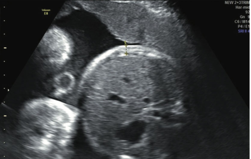
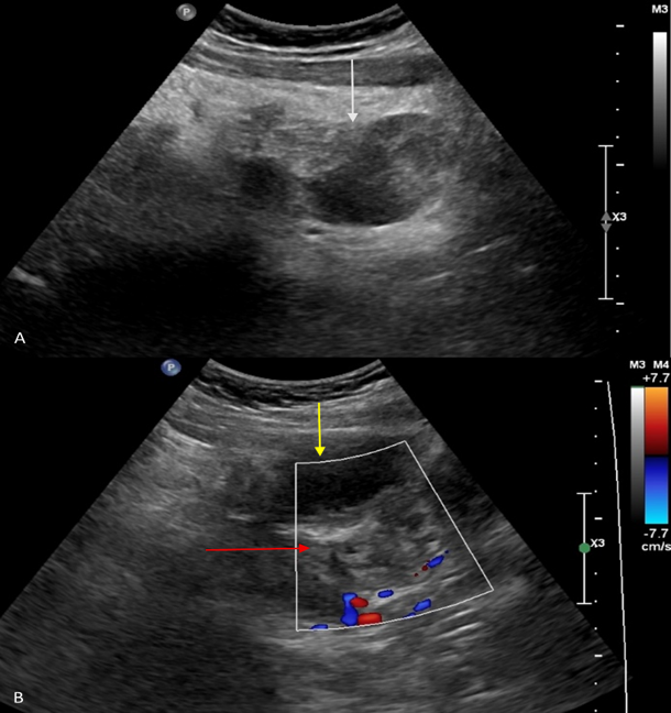
An urgent pelvic ultrasonography demonstrated a well-defined heterogeneous isoechoic lesion measuring 5.4 × 3.7 cm in the right adnexa, with absence of intralesional vascularity [Fig. 1]. The right ovary was not visualized separately, and mild free fluid was noted in the pouch of Douglas. Given persistent acute pain and imaging findings, ovarian torsion was strongly suspected. All routine hematological, biochemical investigations
along with tumor markers were done [Table. 1].
| Category | Test | Date | Result |
|---|---|---|---|
| Hematological | Blood Group | — | B positive |
| CBC | 05/12/25 | Hb: 13.8 g/dL | |
| WBC: 12.9x10³/µL | |||
| Platelets: 2.62 x10⁵/µL | |||
| Urine Culture/ Sensitivity |
06/12/25 | No growth | |
| CUE (Urine Exam) |
06/12/25 | Within Normal Limits | |
| HIV/HBsAg/ HCV/VDRL |
05/12/25 | Non-reactive | |
| Biochemical | T3 | 05/12/25 | 124.12 ng/dL |
| T4 | 05/12/25 | 7.8 ng/dL | |
| TSH | 05/12/25 | 1.96 µIU/mL | |
| Random Blood Sugar |
24/08/25 | 109 mg/dL | |
| Tumor Markers | AFP | 05/12/25 | 1.08 ng/mL |
| CA19-9 | 05/12/25 | 31.67 U/mL | |
| CEA | 05/12/25 | 3.09 ng/mL | |
| CA125 | 05/12/25 | 12.84 U/mL | |
| HE4 | 05/12/25 | 45.8 pmol/L | |
| ROMA Index | 05/12/25 | 6.07% | |
| Beta hCG | 05/12/25 | <5 mIU/mL | |
| LDH | 05/12/25 | 269 U/L |
| S. No. | Timeline | Clinical event & Findings | Implications for “Delayed Intervention” |
|---|---|---|---|
| 1. | 14 years prior | Underwent right ovarian cystectomy | Established a baseline surgical history (No further details were available) |
| 2. | 8 months prior |
Imaging (USG/CT) – outside facility – 4.5 x 3.3 cm right ovarian dermoid cyst. Laparoscopic excision was |
Patient delay in seeking treatment: did not undergo recommended treatment despite experiencing intermittent episodes of abdominal pain |
| 3. | 3 days prior to admission | Onset of acute, progressively worsening pain in the right iliac and suprapubic regions, non-radiating and completing non-responsive to analgesics | Patient delay in seeking acute emergent care: likely irreversible, persistent ischemia |
| 4. | Days of admission |
Tachycardia (P=104/min), localized abdominal tenderness, guarding, and Urgent USG – |
Immediate clinical recognition-suspected ovarian torsion – required emergency exploratory laparotomy |
| 5. | Intraoperatively | 4 x 4 cm blackened, necrotic mass seen completely free-floating (auto-amputated)- an abruptly terminated/absent fallopian tube | This could be due to chronic/intermittent torsion progressed to tissue necrosis and autoamputation |
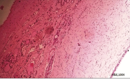
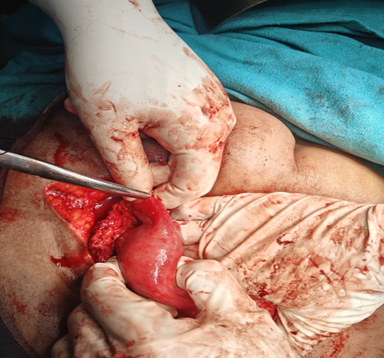
Emergency exploratory laparotomy revealed a solid, well-defined, unruptured mass measuring approximately 4 × 4 cm was found lying free in the pouch of Douglas, retrieved free of adhesions [Fig. 2]. The base of the mass appeared necrotic and blackened, suggestive of an autoamputated dermoid cyst. The right ovary was absent, consistent with chronic torsion and autoamputation, and the right fallopian tube terminated abruptly, possibly consistent with intermittent torsion and chronic ischemia [Fig. 3]. Opportunistic left salpingectomy was performed. The excised mass was removed intact along with left fallopian tube sent for histopathological examination. The postoperative course was uneventful, and the patient was discharged in stable condition on postoperative day 4. On follow up at 3 months, the patient is symptom-free. Histopathological examination confirmed the presence of a dermoid cyst with surrounding severe necrosis (consistent with torsion) and no evidence of malignancy. The timeline of events has been briefly tabulated in [Table. 2].

This case highlights the consequences of delay in intervention for a diagnosed case of ovarian torsion, particularly in the context of mature cystic teratomas.
Autoamputation of the ovarian cyst is defined as cystic masses that are free from tubo-ovarian pedicle and freely mobile within the peritoneal cavity. It is a rare phenomenon and Kennedy et al. were the first to report it[5]. This clinical condition represents the end stage of prolonged or unrecognized adnexal torsion[6].
It is most often identified incidentally during surgical exploration rather than being diagnosed preoperatively. Among the various ovarian pathologies associated with this phenomenon, mature cystic teratoma is the most reported lesion[7]. This is attributed to the high prevalence of dermoid cysts among benign ovarian tumors, slow growing nature and their increased tendency to undergo torsion due to their size, weight, and heterogeneous contents.
The pathogenesis of ovarian autoamputation begins with adnexal torsion, leading initially to venous and lymphatic obstruction followed by arterial compromise. In cases of chronic or intermittent torsion, the necrotic adnexa may gradually detach from its pedicle and become autoamputated. The detached mass may remain free within the peritoneal cavity or adhere to adjacent structures such as the omentum or bowel, occasionally developing a secondary blood supply and behaving as a parasitic mass[8]. This chronic evolution explains the absence of acute symptoms in many patients and contributes to delayed or missed diagnosis.
Preoperative diagnosis of an autoamputated ovarian cyst remains challenging. Ultrasonography may demonstrate a free-floating, partially calcified cystic mass with a fluid debris level, a feature commonly described in autoamputated ovarian cysts[8]. In our case, it demonstrated the absence of blood flow to the mass raising the suspicion of ovarian torsion. The mass was not freely mobile unlike the usual expected radiological finding. However, Doppler ultrasound findings may be misleading, as vascular proliferation within surrounding fibrotic or inflammatory tissue can create a false appearance of vascularity. Consequently, the presence of Doppler flow does not reliably exclude torsion, particularly in cases of chronic or intermittent torsion, contributing to diagnostic delay and progression to autoamputation. In our case, the diagnosis of dermoid cyst was made 8 months back (outside facility) and was advised surgery, but patient did not follow up despite intermittent abdominal pain episodes.
Management depends on the timing of diagnosis and clinical presentation. Once autoamputation has occurred, surgical excision of the detached mass is the treatment of choice. Removal is recommended to prevent potential complications such as infection, adhesion formation, intestinal obstruction, or, rarely, malignant transformation[9]. Histopathological examination is essential to confirm the diagnosis and exclude malignancy.
Laparoscopy is the preferred approach when feasible due to its minimally invasive nature and superior visualization, although laparotomy may be required in cases of large masses or extensive adhesions. In the current study, the patient opted to undergo laparotomy and denied minimally invasive procedure. [Table. 3] mentions the recent case reports highlighting similar presentations with varied symptomatology and lines of management.
| Author name/year | Patient demographics & presentation | Preoperative Findings | Surgical approach & outcomes |
|---|---|---|---|
| Kim et al. 2017 [7] | 34-year-old; history of chronic abdominal pain for 2 years | Smooth, yellowish-white mass -left adnexal origin- no ligamentous support/blood supply. Left ovary/tube completely absent | Laparoscopy; removed via dissection- HPE – mature teratoma; successful pregnancy 7 months later |
| Chaichian et al. 2023 [10] | 46-year-old, presented with secondary amenorrhea for 7 months, experiencing severe dyspareunia but no acute abdominal pain (painless torsion) | Necrotized, oval shaped mass in cul-de-sac; autoamputated right ovary | Laparoscopic successful excision of mass |
| Seyhan et al. 2025 [4] | 33-year-old; presenting with worsening pelvic pain for 3 weeks | 8cm dermoid cyst lodged in cul-de-sac (pouch od douglas), extensively adherent to the bowel and uterus. Left ovary and tube were completely absent | Laparoscopy; the mass was meticulously dissected and removed intact in endobag |
| Unni MM et al. 2025 [11] | Detected at 8 weeks pregnancy; remained asymptomatic ovarian removal was deferred. At term, during cesarean section for cephalopelvic disproportion (CPD), the right ovary was found to be atrophic and was excised. Histopathological examination confirmed ovarian atrophy, consistent with autoamputation |
84 × 74 mm right ovarian cyst noted with history of preterm delivery- planned cerclage and laparoscopic ovarian cystectomy- 12 weeks; A dusky, displaced ovary without attachment was noted attached to omentum (Due to concerns about omental neovascularization, ovarian removal was deferred) |
Later at term- patient under Caesarean section and autoamputated right ovary was retrieved. |
In conclusion, mature cystic teratoma is the ovarian cyst most associated with autoamputation, primarily due to its high propensity for torsion. Ovarian autoamputation represents a late complication of chronic or missed torsion and is rarely diagnosed preoperatively. Preoperative diagnosis of an auto-amputated ovarian cyst is difficult due to nonspecific clinical and imaging signs. Ultrasound may suggest suspicion if a free-floating cyst or absent ovary is seen, while MRI can better define the lesion. Without a clear diagnosis before surgery, minimally invasive planning is limited, often requiring open surgery.
Patient Consent:
Written informed consent was obtained from the patient for publication of this case report.
1. Torsion of huge dermoid cyst in adolescent girl: A case report. Middle East Fertility Society Journal. 2017; 22 (3). Available from: https://doi.org/10.1016/j.mefs.2017.01.003
2. The Auto-Amputated Adnexa: A Review of Findings in a Pediatric Population. Journal of Pediatric and Adolescent Gynecology. 2013; 26 (6). Available from: https://doi.org/10.1016/j.jpag.2012.08.012
3. Ectopic ovary: A case of autoamputated ovary with mature cystic teratoma into the cul‐de‐sac. Journal of Obstetrics and Gynaecology Research. 2007; 33 (3). Available from: https://doi.org/10.1111/j.1447-0756.2007.00538.x
4. Autoamputation of the ovary after missed diagnosis of ovarian dermoid cyst torsion: a case report and review of literature. Journal of the Turkish-German Gynecological Association. 2025; 26 (4). Available from: https://doi.org/10.4274/jtgga.galenos.2025.2025-1-10
5. Amputated calcified ovaries in children.. Radiology. 1981; 141 (1). Available from: https://doi.org/10.1148/radiology.141.1.7291547
6. Spontaneous Autoamputation of Adnexa. CRSLS : MIS Case Reports from SLS. 2024; 11 (3). Available from: https://doi.org/10.4293/crsls.2024.00025
7. A Rare Case of an Autoamputated Ovary with Mature Cystic Teratoma. Journal of Menopausal Medicine. 2017; 23 (1). Available from: https://doi.org/10.6118/jmm.2017.23.1.74
8. Laparoscopic Management of Auto‑Amputated Ovarian Cyst in an Infant. African Journal of Paediatric Surgery. 2026; 21 (1). Available from: https://doi.org/10.67481/ajps.v21i1.998
9. Bilateral Dermoid Ovarian Cysts in a Young Woman – A Case Report and Literature Review. Cancer Diagnosis & Prognosis. 2024; 4 (6). Available from: https://doi.org/10.21873/cdp.10402
10. A Case of Spontaneous Autoamputation of Ovary in a 46-Year-Old Woman: An Uncommon Presentation (Painless Ovarian Torsion) with Unique Diagnostic and Therapeutic Challenges. Case Reports in Medicine. 2023; 2023 Available from: https://doi.org/10.1155/2023/2165226
11. Spontaneous Torsion with Autoamputation of Ovary Presenting in First-trimester as an Adnexal Mass. Journal of South Asian Federation of Obstetrics and Gynaecology. 2025; 17 (S2). Available from: https://doi.org/10.5005/jp-journals-10006-2684
Subscribe now for latest articles and news.