Journal of Medical Sciences and Health
DOI: 10.46347/jmsh.v12.i3.26.34
Year: 2026, Volume: 12, Issue: 3, Pages: 287-292
Original Article
Mouparna Chakraborty 1, P Shashikala 2, Viajaya Kumar B Jatti 3
1Postgraduate Resident, Department of Pathology, S.S. Institute of Medical Sciences & Research Centre, Davangere, Karnataka, India.
2Professor and Head of the Department, Department of Pathology, S.S. Institute of Medical Sciences & Research Centre, Davangere, Karnataka, India.
3Professor and Head of the Department, Department of Forensic Medicine and Toxicology, S.S. Institute of Medical Sciences & Research Centre, Davangere, Karnataka, India.
Address for correspondence: Mouparna Chakraborty, Postgraduate Resident, Department of Pathology, S.S. Institute of Medical Sciences & Research Centre, Davangere, Karnataka, India.
E-mail: [email protected]
Received Date:28 January 2026, Accepted Date:12 July 2026, Published Date:19 August 2026

The spleen is a highly vascular and fragile organ, often vulnerable to rupture following blunt abdominal trauma. However, certain underlying histopathological conditions can predispose the spleen to rupture even with minimal trauma or spontaneously. Therefore, the present study was undertaken to evaluate the gross and histomorphological features of ruptured spleens and identify any associated underlying pathology contributing to splenic rupture. Twenty six specimen of spleen which were resected / sent from autopsy were studied over a six year period. Gross examination focused on rupture grading, size, weight, and capsular integrity. Histopathological examination was done on H&E stained sections and Congo red was used for confirmation of amyloid deposits. Grade IV rupture was the most frequently observed (57.7%), followed by Grade III(23.1%) and V(11.5%). Red pulp congestion and hemorrhage were universally present (100%). Lymphoid hyperplasia was identified in 48% of cases, amyloid deposition in 12%, and neutrophilic perisplenitis in 36%. Fibrinoid necrosis and subcapsular inflammation were also noted in some cases. Histomorphological evaluation of ruptured spleens provides essential insights into both the extent of injury and underlying conditions that may contribute to rupture. This has significant implications for clinical and forensic assessment of spleen trauma.
The spleen is a highly vascular organ that plays a vital role in both hematological and immunological functions, including the filtration of blood, sequestration of aged erythrocytes, and initiation of immune responses. Due to its anatomical location and fragile structure, the spleen is particularly vulnerable to injury during blunt abdominal trauma, such as those sustained in road traffic accidents (RTA)[1-3].
Though splenic rupture is a well-recognized surgical emergency and its clinical diagnosis and surgical management are well-documented, the histomorphological changes associated with splenic rupture remain underexplored in literature[4, 5]. Histopathological evaluation, particularly post-mortem, can uncover underlying pathological processes such as
lymphoid hyperplasia, amyloidosis, or chronic inflammation that may predispose the spleen to rupture[6, 7].
Understanding the microscopic patterns of ruptured spleens has significant clinical, surgical, and medicolegal implications[8]. It helps to differentiate between purely traumatic ruptures and those influenced by pre-existing disease, assists in accurate cause-of-death determination, and improves the pathological classification of splenic injuries[9, 10].
The present study was undertaken in response to this gap. Through a detailed gross and histomorphological analysis of specimen of spleen over a six year period, this study aimed to highlight salient pathological features and compare the findings with previously published literature. In doing so, it seeks to contribute to both forensic histopathology and trauma-based splenic pathology. Aim of the study was to explore the histomorphological changes associated with splenic rupture, to study in detail the gross and microscopic features of ruptured spleens in Hematoxylin and Eosin (H&E) stained sections and use special stains wherever necessary for further evaluation.
This observational study, comprising both retrospective and prospective components, was conducted over a six-year period (2020–2026) in the Department of Pathology. All splenic rupture cases sent for histopathological evaluation during this time were included. The specimens consisted of both autopsy-derived spleens and splenectomy samples obtained following road traffic accidents, clinically diagnosed as splenic rupture, regardless of patient age or sex. Splenectomy done for other causes were excluded.
For retrospective cases, tissue blocks and slides were retrieved from departmental archives and necessary information was obtained from the records. Each specimen underwent detailed gross examination, splenic size, weight, capsular integrity, rupture grade, hemorrhage, and other macroscopic changes were noted. Specimens were then sectioned longitudinally from the hilum to the superior pole at 1 cm intervals (grossing protocol was followed as per the guidelines described in Shameem Shareef’s Manual of Surgical Pathology Grossing Techniques)[10]. Representative bits for study were routinely processed. H&E stained sections were studied, Congo red stain was done in which amyloidosis was suspected. On H&E all microscopic slides and gross samples were examined and reviewed by senior pathologists.
Findings were documented, grouped based on rupture grading, and correlated with histopathological features. All results were compiled and analyzed. Ethical clearance for the study was obtained from the Institutional Ethics Committee, and proper anonymization protocols were followed for use of archived autopsy material.
A total of 26 ruptured spleen were analyzed, of which 22 (84.6%) belonged to males and 4 (15.4%) females, with a male-to-female ratio of 5.5:1. The most common age group was 31–50 years (10 cases, 38.4%), followed by 21–30 years (7 cases, 26.9%), 51–70 years (5 cases, 19.2%).
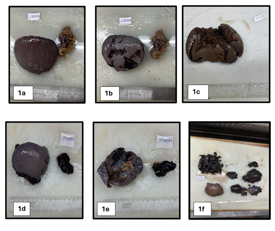
All cases had a history of blunt abdominal trauma sustained in road traffic accidents (RTA). Among the 26 specimens, 19 (73.1%) were obtained from autopsies and 7 (26.9%) were post-operative splenectomy specimens. Gross examination revealed capsular rupture in all cases, with variable degrees of hemorrhage. Grade IV rupture was the most common, seen in 15 cases (57.7%), followed by Grade III in 6 cases (23.1%), Grade V in 3 cases (11.5%), and Grade II in 2 cases (7.7%) ([Fig. 1] a-f).
| Sl. No. | Size (in cms) | Gross Findings | Microscopy |
|---|---|---|---|
| 1 | 7x5.5x3 | Congestion and haemorrhage. | Hemorrhage, Lymphoid follicle hyperplasia |
| 2 | 9x6x3 | Inferomedial border laceration and haemorrhage. | Hemorrhage, Lymphoid follicle hyperplasia |
| 3 | 11.5x7.5x4 | Anterior surface laceration and haemorrhage. | Hemorrhage, homogenous eosinophilic deposits in arterioles |
| 4 | 12x9.5x5 | Focal area of haemorrhage. | Hemorrhage, Lymphoid follicle hyperplasia |
| 5 | 7x6x4 | Breach of capsule and haemorrhage. | Hemorrhage, Neutrophilic infiltrate at the ruptured area |
| 6 | 12x6x4 | Breach of capsule and haemorrhage. | Hemorrhage, Neutrophilic infiltrate at the ruptured area |
| 7 | 11.5x7x5.5 | Anterior and posterior surface laceration and haemorrhage. | Hemorrhage, homogenous eosinophilic deposits in arterioles |
| 8 | 10x6x4 | Breach of capsule and haemorrhage. | Hemorrhage, Neutrophilic infiltrate at the ruptured area |
| 9 | 8x6x4 | Breach of capsule and haemorrhage. | Hemorrhage, Lymphoid follicle hyperplasia |
| 10 | 12x8x4 | Breach of capsule and haemorrhage with perisplenitis. | Hemorrhage, Lymphoid follicle hyperplasia |
| 11 | 7x5x3 | Congestion and haemorrhage. | Hemorrhage, Neutrophilic infiltrate at the ruptured area |
| 12 | 14x5x3 | Posterior tear and haemorrhage. | Hemorrhage, homogenous eosinophilic deposits in white pulp |
| 13 | 10x7x2 | Congestion and haemorrhage. | Hemorrhage, Lymphoid follicle hyperplasia |
| 14 | 11x7x3 | Posterior surface laceration and haemorrhage. | Hemorrhage, Neutrophilic infiltrate at the ruptured area |
| 15 | 14x9x3.5 | Haemorrhage. | Hemorrhage, Neutrophilic infiltrate at the ruptured area |
| 16 | 14x9x6 | Anterior laceration and haemorrhage. | Hemorrhage, Lymphoid follicle hyperplasia |
| 17 | 9.5x5x4 | Haemorrhage. | Hemorrhage, homogenous eosinophilic deposits in white pulp |
| 18 | 9x7x3.5 | Breach of capsule and haemorrhage. | Hemorrhage, Lymphoid follicle hyperplasia |
| 19 | 7x5.5x3 | Breach of capsule , Congestion and haemorrhage. | Hemorrhage, Lymphoid follicle hyperplasia |
| 20 | 9x5x4 | Congestion and haemorrhage. | Hemorrhage, Lymphoid follicle hyperplasia |
| 21 | 11x7x3 | Congestion and haemorrhage. | Hemorrhage, Lymphoid follicle hyperplasia |
| 22 | 8x6x4 | Congestion and haemorrhage. | Hemorrhage, Neutrophilic infiltrate at the ruptured area |
| 23 | 10.5x6x3 | Congestion and haemorrhage. | Hemorrhage, Lymphoid follicle hyperplasia |
| 24 | 8x5x4 | Congestion and haemorrhage. | Hemorrhage, Neutrophilic infiltrate at the ruptured area |
| 25 | 9x6x3 | Congestion and haemorrhage. | Hemorrhage, Lymphoid follicle hyperplasia |
| 26 | 9.5x5x4 | Congestion and haemorrhage, anterior surface laceration. | Hemorrhage, Neutrophilic infiltrate at the ruptured area |
Spleen weights ranged from 90–250 grams. Weight of 22 spleens (84.6%) were within normal limits (90–140 g), 3 (11.5%) were mildly increased (150–200 g), and 1 case (3.9%) showed significant increase (250 g).
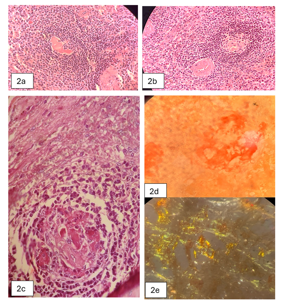
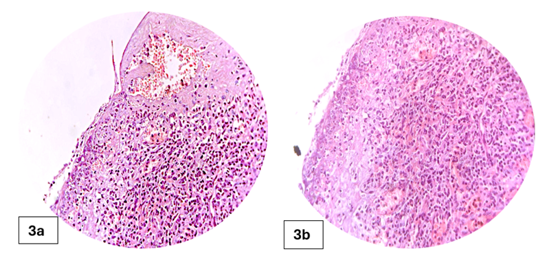
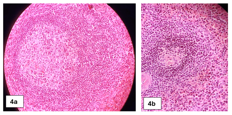
On microscopy, all 26 cases (100%) showed red pulp congestion and hemorrhage at the rupture site. Additional histopathological findings included [Table. 1] were lymphoid hyperplasia ([Fig. 4] a,b) in 13 cases (50%), amyloid deposition ([Fig. 2] a-e) in 3 cases (11.5%), neutrophilic perisplenitis in 9 cases (34.6%), subcapsular neutrophilic infiltrates ([Fig. 3] a,b) in 4 cases (15.4%), fibrinoid necrosis in 2 cases (7.7%),
perisplenic inflammation in 2 cases (7.7%) and no significant pathology 4 cases (15.4%).
The spleen is one of the most frequently injured solid organs in blunt abdominal trauma, accounting for up to 42% of such injuries[1]. Its anatomical position, friable capsule, and rich vascularity make it particularly prone to rupture in high-impact injuries such as road traffic accidents (RTA).
In our study, 100% of the cases (n = 26) were associated with blunt trauma due to RTA, consistent with findings by Fomin et al.[2] in their post-mortem analysis of traumatic splenic injuries.
A marked male predominance (84.6%) was observed, which corresponds with the findings of Al-Kindi et al.[9], who noted that young to middle-aged males were at highest risk which is probably attributed to increased occupational exposure and usage of vehicles in this age group.
Grade IV ruptures were most frequent (57.7%) in our series, reflecting the severity of trauma encountered. This observation aligns with Farhi and Ashfaq[3], who documented that higher-grade ruptures are common in traumatic spleens. Interestingly, 84.6% of spleens weighed within the normal range, supporting earlier assertions that splenomegaly is not essential for rupture. However, Parsons et al.[4] noted that even mild enlargement may predispose the spleen to injury under pressure.
Microscopically, all cases (100%) showed red pulp congestion and hemorrhage. Lymphoid hyperplasia was present in 50% of cases ([Fig. 4] a,b), comparable to reports by Farhi et al.[3], indicating underlying immune activation. In 11.5% of cases, amyloid deposits were identified by Congo red staining. This finding resonates with Renzulli et al.[5], who highlighted that amyloid infiltration compromises tissue elasticity and may predispose to spontaneous rupture, even in trauma-dominant settings.
Other histological features included neutrophilic perisplenitis (34.6%), subcapsular inflammation (15.4%), and fibrinoid necrosis (7.7%). These patterns suggest a spectrum of acute and chronic inflammatory responses. Dalrymple et al.[6] emphasized the importance of such histologic evidence in distinguishing between pre-existing disease and trauma-induced changes, especially in forensic practice.
Presence of perisplenic inflammation in one case aligns with the insights of Byard et al.[7], who proposed that capsular thickening or adhesions may hint at silent, prior trauma or ongoing low-grade inflammation. This reinforces the value of thorough histopathological examination in both clinical and medico-legal contexts.
This comparative analysis confirms that our results are in line with existing literature, while also highlighting the importance of histomorphology in detecting predisposing conditions especially in autopsy cases where aetiology needs to be established for legal and clinical reporting.
Limited Sample Size and Single-Centre Design. Although the study included 26 cases over six years, the sample size remains modest for drawing broad epidemiological conclusions. Being conducted in a single tertiary-care centre may also limit generalizability to other populations or trauma settings.
Lack of Statistical Analysis. Due to the descriptive nature of the study, no advanced statistical correlations were performed. Potential associations between histological features and clinical variables (e.g., age, rupture grade) could not be statistically validated.
Retrospective Data Retrieval Bias. Some cases (before 2023) were retrospective, and relevant clinical history or injury details were incomplete or missing in archived records, potentially influencing clinicopathological correlation.
Lack of Clinical Follow-Up. As most specimens were derived from autopsies or emergency splenectomies, patient follow-up data was unavailable. Therefore, correlations with prognosis, outcomes, or recurrence could not be assessed.
Histomorphological evaluation of ruptured spleens offers critical insights beyond gross examination alone. In this study, all cases were associated with blunt trauma following road traffic accidents, with red pulp congestion and hemorrhage being universal findings. However, the identification of additional features such as lymphoid hyperplasia, amyloid deposition, and perisple- nitis underscores the potential role of underlying pathological conditions in contributing to splenic fragility.
These findings highlight the importance of detailed microscopic examination in distinguishing between purely traumatic ruptures and those complicated or predisposed by pre-existing disease. Such differentiation is essential not only for accurate clinical management, but also for forensic investigations, where establishing the cause and context of rupture can have legal implications.
The study reinforces that routine histopathological analysis should be considered a vital step in the evaluation of all ruptured spleens — especially in autopsy practice — to ensure that hidden disease processes are not overlooked.
Conflict of Interest:
The authors declare no conflict of interest related to this study.
Funding:
This research received no external funding and was conducted as part of routine departmental academic work.
Acknowledgements:
The authors would like to express sincere gratitude to the Department of Pathology S.S. Institute of Medical Sciences and Research Centre, for providing access to archival material and laboratory support. Special thanks to Dr. Shashikala P, Head of Department, for her continuous guidance and encouragement throughout the study.
1. Kraus MD, Kim KC, DeMay RM. Histopathology of splenic trauma. Human Pathology. 2003;34(5):500–5.
2. Euler SA, Schindera ST. Postmortem imaging and autopsy correlation of blunt abdominal trauma: a forensic perspective. Journal of Forensic Radiology and Imaging. 2017;8:20–5.
3. Farhi DC, Ashfaq R. Histological evaluation of traumatic splenic ruptures: patterns and pitfalls. American Journal of Clinical Pathology. 1996;105(4):445–50.
4. Parsons RJ, Thompson WE. The significance of splenic size in trauma. Surgery gynecology & obstetrics. 1958;106(6):705–12.
5. Systematic review of atraumatic splenic rupture. British Journal of Surgery. 2009; 96 (10). Available from: https://doi.org/10.1002/bjs.6737
6. Dalrymple CJ, Greenson JK. Distinguishing trauma-induced vs. pre-existing changes in ruptured spleens: a histologic review. Forensic Science International. 2001;123(2–3):147–52.
7. Byard RW, Simpson E. Forensic issues in splenic rupture: a review of mechanisms and histologic clues. Forensic Science, Medicine and Pathology. 2011;7(1):36–40.
8. O’Malley RB, Mellnick VM. Systemic conditions and the spleen: radiologic-pathologic correlation. In: O’Malley RB, Mellnick VM, editors. Radiologic-Pathologic Correlations from Head to Toe. Springer; 2012. p. 219–29
9. Al-Kindi H, Devi L, George M. Splenic pathology in traumatic rupture of the spleen: a five-year study. Oman Medical Journal. 2009;24(3):210–13.
10. Shareef S. Manual of Surgical Pathology Grossing Techniques. 1st ed. Hyderabad: Paras Medical Publishers; 2019.
Subscribe now for latest articles and news.