Journal of Medical Sciences and Health
DOI: 10.46347/jmsh.v12.i3.25.241
Year: 2026, Volume: 12, Issue: 3, Pages: 230-238
Original Article
Tarun Kumar D Jain 1, Sanjana A S 2, Rajendra Okade 3
1 Junior Resident, Department of Dermatology, Venereology and Leprosy, BGS GIMS Hospital, Bengaluru, Karnataka, India.
2Professor and Head of Department, Department of Dermatology, Venereology and Leprosy, BGS GIMS Hospital, Bengaluru, Karnataka, India.
3Professor, Department of Dermatology, Venereology and Leprosy, BGS GIMS Hospital, Bengaluru, Karnataka, India.
Address for correspondence: Tarun Kumar D Jain, Junior Resident, Department of Dermatology, Venereology and Leprosy, BGS GIMS Hospital, Bengaluru, Karnataka, India.
E-mail: [email protected]
Received Date:30 June 2025, Accepted Date:04 June 2026, Published Date:19 August 2026

Background: Chronic Spontaneous Urticaria (CSU) is very common dermatological condition characterised by repeated episodes of wheals lasting for ≥ 6 weeks. Metabolic syndrome could be present with the same etiopathogenesis and urticarial markers like CRP, D-dimer, prolonged PT and aPTT are elevated which are likely to be associated with severity and prognosis of Urticaria. Metabolic syndrome has been discovered to be related to Chronic Spontaneous Urticaria.
Aims: 1) To estimate the metabolic syndrome among patients with Chronic Spontaneous Urticaria. 2) To estimate the biomarkers among patients with Chronic Spontaneous Urticaria. 3) To describe demographic profile of patients with Chronic Spontaneous Urticaria.
Methods: A Cross-sectional study for 1 year of 18 to 60 yrs. Diagnosis of metabolic Syndrome was based on the National Cholesterol Education Programme (NCEP) Adult Treatment Panel III. FBS, CRP, D-dimer, Lipid Profile, PT, aPTT were measured.
Results: Total of 83 cases of Chronic Spontaneous Urticaria were enrolled. 32(38.6%) patients were males, and 51(61.4%) patients were females with chronic spontaneous urticaria. 14 out of 32 male patients and 28 of 51 female patients of CSU developed metabolic syndrome. 50% Males and 40.07% Females with elevated D-Dimer developed metabolic syndrome with a significant P value of (P=0.02). 33.33% male patients and 57.14% female patients with raised CRP developed metabolic syndrome with a significant value of 0.03.
Conclusion: Chronic Spontaneous urticaria patients with elevated Urticarial Biomarkers can serve as marker for cardiovascular risks including metabolic syndrome, hypertension, obesity and hyperlipidemia and could be associated with severity of Urticaria and Prognosis.
Chronic urticaria (CU) is defined by recurrent wheals lasting for a minimum of 6 weeks. CU can be classified into two primary groups based on the presence of lesions: those that occur spontaneously, known as chronic spontaneous urticaria (CSU), and those which are triggered by physical or environmental factors, termed chronic inducible urticaria (CIndU)[1].
Urticaria impacts a significant portion of the population, with estimates suggesting that 15-20% experience it at
least once or more in their lifetime. CSU is estimated to impact 0.5%-1% of adults, substantially diminishing their quality of life[1]. Females experience this condition more frequently than males, with the majority of patients falling between the ages of 20 and 40 years, with an average age of 33 years[2]. About 30% of patients frequently experience recurrences lasting for months or even years[3].
While various factors such as food allergies, emotional disturbances, intolerance to food additives, and chronic infections are considered potential causes of the disease,[4] its exact underlying cause remains elusive for many patients despite thorough investigations.
Various lines of evidence suggest that different biological systems, such as immunity, inflammation, and coagu- lation, could collectively contribute to a shared mechanism resulting in the formation of wheals.
Autoimmunity plays a significant role in the pathophysiology of chronic urticaria, as evidenced by wheal and flare reactions induced by autologous serum injections, and the identification of several autoantibodies such as anti-IgE in affected patients. However, these antibodies are detectable in only one-third of chronic urticaria cases, and the autologous serum skin test (ASST) fails to provoke a wheal-flare reaction in approximately half of them[5-7]. Hence, other factors likely contribute to the development of chronic urticaria.
Recent evidence suggests that additional patho mechanisms may play a role, such as alteration of intracellular signalling pathways in basophils and mast cells, abnormal innate immunity responses, and the concurrent activation of inflammatory and coagulation systems[8-10]. Eosinophils, by expressing tissue factor and activating the extrinsic coagulation pathway, facilitate the conversion of prothrombin to thrombin through activated factor X, leading to the generation of Platelet fragments[11, 12]. This process contributes to urticaria by enhancing vascular permeability and plasma extravasation, subsequently triggering degranulation of skin mast cells[13-15]. Furthermore, thrombin's action on fibrinogen results in the formation of fibrin, which is eventually degraded by plasmin, producing fibrin degradation products and plasma D-dimer. These molecules serve as potential biomarkers for assessing disease severity in both acute and chronic urticaria.
C-reactive protein, a commonly used inflammatory response marker, is associated with Chronic Spontaneous Urticaria activity[16]. Patients with chronic spontaneous urticaria (CSU) exhibit elevated levels of oxidative stress, which occur concomitantly with systemic inflammation[17]. A linear relationship in the concentration of the thrombotic marker D-dimer was detected in Metabolic syndrome (MetS), in association with chronic inflammation and oxidative stress[18].
The aim of the present study was to estimate the prevalence of MetS and biomarkers in patients with CSU. Due to lack of sufficient data and studies in our country about possible link between metabolic syndrome and urticarial biomarkers in chronic spontaneous urticaria, conducting this study is necessary for establishing such an association.
Objectives of the Study:
1. To estimate the metabolic syndrome among patients with Chronic Spontaneous Urticaria.
2. To estimate the biomarkers among patients with Chronic Spontaneous Urticaria.
3. To describe demographic profile of patients with Chronic Spontaneous Urticaria.
Study design:
A hospital based cross-sectional observational study was conducted among patients clinically diagnosed with Chronic Spontaneous Urticaria visiting the Department of Dermatology, Venerology, Leprosy, from May 2023 to May 2024.
Study duration:
The study was conducted over a period of 12 months from May 2023 to May 2024. This duration comprised of protocol development, research and ethics committee approval, data collection, analyses of the data and report writing.
Study participants:
Male patients having Chronic Spontaneous Urticaria in the age group of 18-60 years presenting to the Dermatology Department from May 2023 to May 2024.
Sampling technique:
Male patients having Chronic Spontaneous Urticaria in the age group of 18-60 years presenting to the Department of Dermatology, were selected purposively in the study.
Inclusion criteria:
All patients who are aged between 18-60yrs with clinically diagnosed as CSU.
Patients on antihistamines after a washout period of 3 days, were included in the study.
Exclusion Criteria:
Patients with inducible urticaria, urticarial vasculitis, acute urticaria.
On medications (aspirin, clopidogrel, warfarin, heparin, omalizumab, steroid, immunosuppressants) that could influence the course of urticaria or coagulation/fibrinolysis pathway.
Known case of Diabetes, Hypertension and are on oral hypoglycaemics, antihypertensives or lipid lowering agents were excluded from the study.
Pregnant/lactating women were excluded from the study.
Study procedure:
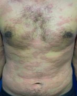
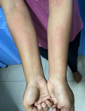
All patients with CSU meeting the inclusion criteria were enrolled into the study. Informed consent of the enrolled patients was taken. A pre structured proforma was used to collect the demographic data. A detailed history was taken and a thorough cutaneous and systemic examination was done. Height, weight, BMI were recorded. Clinical photographs were taken with patients consent. Diagnosis of MetS was based on the national cholesterol education Programme (NCEP) adult treatment panel III by the presence of 3 or more of the following criteria.
Relevant investigations were sent and reports were recorded.
waist circumference ≥ 102 cm (40 inches) in men or ≥ 88 cm (35 inches) in women;
hypertriglyceridemia (triglyceride level ≥ 150 mg/dl);
high-density lipoprotein (HDL) cholesterol level < 40 mg/dl in men or < 50 mg/dl in women;
blood pressure ≥ 130 ⁄85 mmHg;
fasting plasma glucose level ≥ 100 mg/dl.
Venous samples were taken after an overnight fast and abstinence from vigorous activity for 24 h. Fasting glucose, total cholesterol, HDL cholesterol, and triglyceride levels were measured by GPO-PAP/enzymatic calorimetric end point method. Abdominal circumference was measured using a flexible measuring tape to the nearest centimetre, 2cm above the navel, with the patient in standing position. Blood pressure was measured with the patient in sitting position. Serum concentrations of CRP were measured by immunoturbidometric method. Coagulation parameters such as prothrombin time (PT), and activated partial thromboplastin time (aPTT) were assayed in all study subjects. Plasma D-dimer levels were estimated quantitatively in all patients. Plasma D-dimer levels ≥ 500ng/mL were considered elevated (normal levels ≤500 ng/mL as per manufacturer's manual). The Statistical software namely SPSS 25.0, and R environment ver.3.2.2 were used for the analysis of the data and Microsoft word and Excel have been used to generate graphs, tables etc.
Ethical considerations:
The approval to conduct the study was sought from the Research monitoring committee and Institutional Ethics Committee (IEC) with the reference number: IEC/App/Apr/2023/020. Informed consent was obtained from the study participants before data collection. Informed consent was taken by the investigator, after explaining the purpose of the study. The freedom to withdraw from the study at any time during the interview was also explained prior to taking of the informed consent. Data were analyzed in aggregate and access to the collected data was limited only to me, my guide and co-guide.
Sample Size of Estimation:
83 cases of Chronic Spontaneous Urticaria attending to the Department of Dermatology out- patients during the study period of 1 year.
A total of 83 cases of Chronic Spontaneous Urticaria were enrolled. Mean age of male population (Mean ± SD) was 35.65±12.93 and mean age of female population (Mean ± SD) was 35.80±12.95 [Table. 1], [Fig. 1].
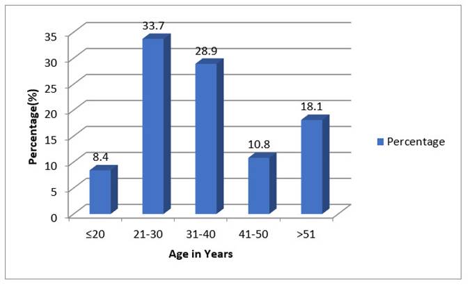
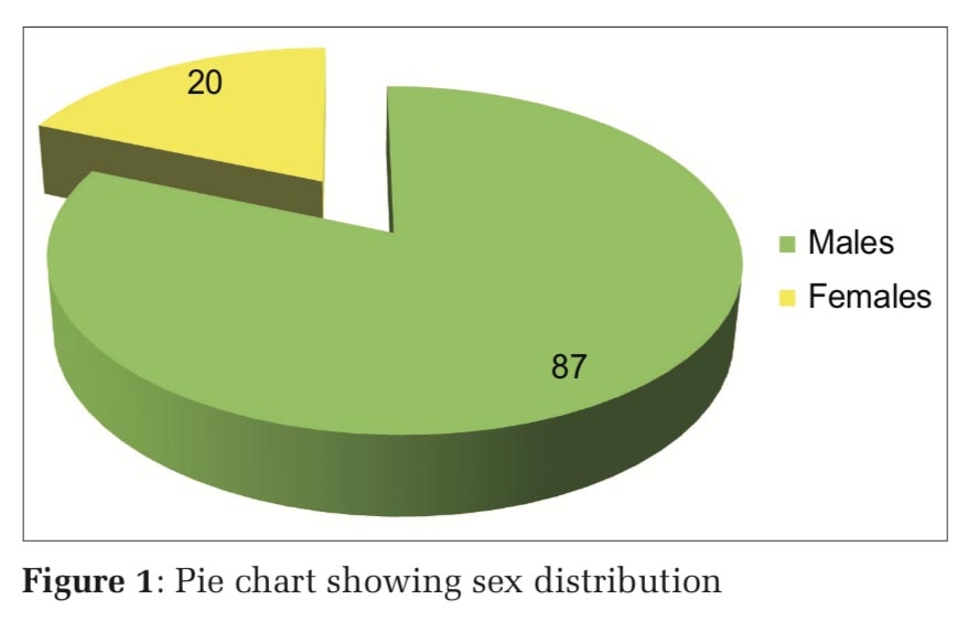
Majority of our patients (33.7%) belonged to 21 to 30 years of age with a mean age of 35.65±12.93. Thirty-two (38.6%) patients were males, and 51(61.4%) patients were females with chronic spontaneous urticaria [Table. 2]. Fourteen (14) out of 32 male patients of CSU developed metabolic syndrome (43.75%). Twenty-eight (28) of 51 female patients of CSU developed metabolic syndrome (54.9%) [Table. 3]. One Male patient out of 2 with elevated waist circumference had metabolic syndrome. Seventeen female patients out of 23 with elevated waist circumference had metabolic syndrome with a significant P value <0.001 [Table. 10].
|
Age in years |
No. of Patients (n= 83) |
Percentage (%) |
|---|---|---|
|
≤20 |
7 |
8.4 |
|
21-30 |
28 |
33.7 |
|
31-40 |
24 |
28.9 |
|
41-50 |
9 |
10.8 |
|
>51 |
15 |
18.1 |
Majority of our patients (33.7%) belonged to 21 to 30 years of age with a mean age of 35.65±12.93.
|
Gender |
No. of Patients |
Percentage (%) |
|---|---|---|
|
Male |
32 |
38.6 |
|
Female |
51 |
61.4 |
|
Total |
83 |
100.0 |
Majority of the Chronic spontaneous urticaria patient were females (n= 51,61.4%) as compared to males (n=32,38.6%)
|
Metabolic |
No of |
Percentage |
|---|---|---|
|
Male |
14 |
33.33 |
|
Female |
28 |
66.67 |
|
Total |
42 |
100.00 |
Female patient (n=28, 66.67%) had developed metabolic syndrome as compared to males (n= 14,33.33%)
|
PT (Prothrombin |
No. of |
Percentage |
|---|---|---|
|
Male |
2 |
40.00 |
|
Female |
3 |
60.00 |
|
Total |
5 |
100.0 |
3 female patients (60%) of metabolic syndrome had prolonged PT as compared to only 2 male patient (40%).
|
D-dimer |
No. of |
Percentage |
|---|---|---|
|
Male |
7 |
46.67 |
|
Female |
8 |
53.33 |
|
Total |
15 |
100 |
53.33% of female patients of metabolic syndrome had elevated D-dimer as compared to male patients (46.67%)
|
C-Reactive |
No. of |
Percentage |
|---|---|---|
|
Male |
1 |
11.11 |
|
Female |
8 |
88.89 |
|
Total |
9 |
100.00 |
8 female patients of metabolic syndrome (88.89%) had elevated C-Reactive Protein and 1male patients of metabolic syndrome (11.11%) had elevated C-Reactive Protein.
|
CRP and D-dimer |
Male (n) |
Percentage (%) |
Female (n) |
Percentage (%) |
|---|---|---|---|---|
|
Elevated |
0 |
0 |
6 |
14.28 |
The study revealed that 14.28 % patients of metabolic syndrome had elevated CRP and D-dimer both
|
PT and APTT |
Male (n) |
Percentage (%) |
Female(n) |
Percentage (%) |
|---|---|---|---|---|
|
Prolonged |
0 |
0 |
2 |
(PT= prothrombin time, APTT= activated partial thromboplastin time). The study revealed that 4.76 % patients of metabolic syndrome had prolonged PT and APTT both.
|
Gender (M/F) |
Correlation coefficient |
P value |
|---|---|---|
|
Waist Circumference |
0.023 |
0.86 |
|
BP |
0.06 |
0.72 |
|
FBS |
0.182 |
0.25 |
|
TGs |
0.378 |
0.02- significant |
|
HDL |
0.115 |
0.47 |
|
PT |
-0.122 |
0.44 |
|
aPTT |
0.001 |
0.99 |
|
D Dimer |
0.362 |
0.03- significant |
|
CRP |
0.381 |
0.02- significant |
(BP= blood pressure, FBS= fasting blood glucose, TGs= triglycerides, HDL= high density lipoprotein, PT= prothrombin time, APTT= activated partial thromboplastin time, CRP= C reactive protein). Patients with metabolic syndrome were associated with elevated CRP and D-dimer with a significant P value. Patients of metabolic syndrome had elevated triglycerides levels with a significant P value.
All 8 male patients with elevated FBS had metabolic syndrome. Nine female patients out of 12 with elevated FBS had metabolic syndrome. Seven male patients out of 14 with elevated D-dimer developed metabolic syndrome and 8 female patients out of 17 with elevated D-dimer developed metabolic syndrome with a significant P value of 0.02 [Table. 5]. One male patient out of 3 with elevated CRP developed metabolic syndrome and eight female patients out of 14 with elevated CRP developed metabolic syndrome with a significant value of 0.03 [Table. 6]. Fourteen (14) male patients and 22 female patients of metabolic syndrome had elevated blood pressure. Two male patients and 3 female patients of metabolic syndrome had prolonged PT with a significant P value of 0.04 [Table. 4]. One male patient and 9 female patients of metabolic syndrome had prolonged aPTT and 10 male patients and 15 female patients of metabolic syndrome had elevated triglycerides. Nine male patients and 25 female patients of metabolic syndrome had reduced HDL levels with a significant P value of 0.04. 14.28 % patients of metabolic syndrome had elevated CRP and D-dimer both [Table. 7]. 4.76 % patients of metabolic syndrome had prolonged PT and aPTT both [Table. 8].
|
Gender (M/F) |
Correlation coefficient |
P value |
|---|---|---|
|
Waist Circumference |
0.56 |
<0.001- significant |
|
PT |
-0.289 |
0.04- significant |
|
aPTT |
-0.240 |
0.07 |
|
D Dimer |
-0.32 |
0.02- significant |
|
CRP |
0.30 |
0.03- significant |
|
FBS |
0.052 |
0.74 |
|
Lipid (TGs) |
0.211 |
0.18 |
|
HDL |
0.30 |
0.04- significant |
17 female Patients of metabolic syndrome had an elevated waist circumference with a significant P value (< 0.001). 3 female patients of metabolic syndrome had prolonged prothrombin time (PT) with a significant P value (0.04). 8 female patients of metabolic syndrome had an elevated D-dimer with a significant P value (0.02). 8 female patients of metabolic syndrome had CRP with a significant P value (0.03). 25 female patients of metabolic syndrome had a reduced HDL levels with a significant P value (0.04).
The association between chronic spontaneous urticaria and metabolic syndrome has been focused of many previous studies. However, the findings were variable. The purpose of our study was to estimate whether metabolic syndrome was present in individuals with chronic spontaneous urticaria and to evaluate the metabolic syndrome parameters in relation to it and to see whether there is elevated urticarial biomarkers in these patients that could be used as a marker of an underlying systemic condition that could be treated if detected early.
In our study 83 cases diagnosed clinically as chronic spontaneous urticaria were investigated for conditions like hypertension, hyperlipidaemia, fasting blood glucose levels. They were subjected to blood investigations for measuring HDL levels, triglycerides levels and fasting blood sugar levels and anthropometric measurements like waist circumference.
In our study majority of the patients (33.7%) belonged to 21 to 30 years of age with a mean age of 35.65±12.93 which was similar to the study by Chauhan et al where the age group was between 18-40years[19]. This is due to the disease as itching promotes patient to seek early health care professionals.
CSU was more commonly seen in female patients (61.4%) than male patients (38.6%) in our study, which is similar to findings seen in several studies[20-23]. Also, females experience higher incidence and severity, leading to extended disease durations and a more significant impact on their overall wellbeing compared to men. This difference is due to elevated estradiol levels during fertile phase of women, leading to mast cell activation.
14 out of 32 males (43.75%) and 28 out of 51 females (54.9%) of CSU patients had metabolic syndrome. In our study around 50% patients had developed metabolic syndrome which is in contrast to a study by Young-min et al. where 30% of participants developed it and majority of them were males[24]. This higher prevalence is due to early age of diagnosis of our patients.
In study by Gupta et al.[25] female patient of CSU with metabolic syndrome had elevated waist circumference corresponding to central obesity, which is similar to our study where female was majority with elevated waist circumference with a significant P value (<0.001).
Females are to a greater extent prone to auto immune diseases and obesity due to hormonal imbalances and irregular cycles attributing to hidden component of MetS. Also, it gets early diagnosed in females as women in our society are more often reduced to their appearance and therefore experience more external pressure. On the other hand, there is also a different understanding of illness between men and women in our society, which could lead to these items being of greater importance to women.
All male patient with elevated FBS had developed metabolic syndrome, whereas 9 out of 12 females had elevated FBS. Males are prone to increased insulin resistance among metabolic syndrome due to elevated TNF -α, a powerful regulator of the synthesis of IL-6, leading to impaired glucose tolerance.
In our study, 9 male patient and 25 female patients of metabolic syndrome had reduced HDL levels with a significant P value of 0.04. Reduced HDL levels are associated with increased risk of cardiovascular events and mortality as by study by yang et al.[26]. Therefore, early detection of this parameter could also lead to early intervention in treating CSU patient and preventing morbidity and mortality. 10 male patients and 15 female patients of metabolic syndrome had elevated triglycerides in our study. A similar relation was elicited by a study using a population-based dataset in Taiwan, involving 9798 adults with CSU and controls[27].
A relationship between CSU and serum lipids and fatty acids was previously suggested by Kobayashi,[28] who hypothesized the role of omega- 6 and omega-3 series of polyunsaturated fatty acids and lipid peroxidation as mediators in CSU. Now this finding is relevant to metabolic syndrome or not is difficult but further studies could pave way in finding such an association. Increased triglycerides levels in patient with CSU patient, leading to increased inflammatory cytokines and increased biomarkers could be a reason for this finding in our study.
14 male patient and 22 female patients of metabolic syndrome had elevated blood pressure in our study. The association between CSU and hypertension was examined by a retrospective cohort study of 2,460 patients with CSU and 9,840 age-, sex-, and index year-matched control patients, using the National Health Insurance of Taiwan database. Patients with CSU were found to have a 1.37-fold higher risk of developing subsequent hypertension than the non-CSU cohort after adjusting for sex, age, comorbidities.
Hypertension could be due to elevated IL-6,10 with elevated TNF-alpha levels among urticarial patients. Whether this finding is directly associated with CSU or a pre-existing hidden co morbid condition or not could not be found with a statistical value. Among 52 females 17 had elevated D-dimer levels with 8 (47.05%) of them developing metabolic syndrome. Among 32 males, 14 had elevated D-dimer levels with 7 (50%) of them developing metabolic syndrome, which is similar to the Brazilian study[29] where there was a strong and positive relationship between elevated D-dimer and CSU among metabolic syndrome patient. It is a marker of fibrinolysis activation among individuals with metabolic syndrome. MetS patient have low level of chronic inflammation characterised by increased cytokines and coagulation factors.
Though we are unable to measure CSU severity due to study design but there is a strong correlation with these variables. This biomarker was significantly associated with a P value of (0.02). CRP is a marker of systemic inflammation. Among metabolic syndrome patient there is a low level of systemic inflammation with elevated IL-10, which is interplaying with other proinflammatory cytokine in bringing the persistence of urticaria. The elevated CRP levels found in our study point towards a systemic inflammatory response in CU. Whether this is a mere association or has a role in the pathogenesis is not clear. CRP, being an acute phase reactant, may enhance urticarial inflammation and thus disease activity in CU.
In our study 1 out of 3 male patient and 8 out of 14 female patients had elevated CRP levels among patient with MetS with a significant P value (P-0.03) which is similar to a study by Kolkhir et al.[30]. 14.28% patient had both elevated CRP and D-dimer levels in metabolic syndrome patients.
In a Brazilian study,[29] though both CRP and D-dimer were evaluated among CSU patients. D-dimer was directly related to CSU patient and with severity of CSU. No such direct correlation was made with elevated CRP levels. Therefore, evaluating it among CSU patient would be beneficial in assessing the severity and planning treatment.
In our study 2 female patient (4.76%) had both PT, aPTT prolonged with a significant P value of 0.04 which is similar to study done by Sharmeen et al.[31] where these parameters were elevated. PT and aPTT are markers of coagulation cascade in patients of CSU. Inflammation activates coagulation, and coagulation provokes the inflammatory system. This inflammation is present in patient with metabolic syndrome. Therefore, finding a direct link is difficult and yet a large cohort study is required in finding this association. Various studies have found separately the association of metabolic syndrome with coagulation markers, MetS with D-dimer and CRP but no such study is there to find a correlation among all these biomarkers in a study population. Our study helps in finding this association.
Limitations:
Smaller sample size owing to limited time period. Further multicentric studies with larger sample size across the subcontinent will help us to determine the association between MetS, CSU and elevated CRP, D-dimer levels, coagulation profile (PT, aPTT). Treatment outcome measures were not part of the study.
CSU shares some pathobiological pathways with MetS, including a pro-inflammatory state, increased oxidative stress, alterations in adipokine profile and activation of the coagulation system. The role of obesity seems to be of interest. Further research is required to assess the association with MetS and its practical implications in terms of prognosis and treatment response among CSU patients. The exact role of D-dimer in predicting disease activity need to be elucidated further after large systematic studies and long-term post treatment follow up. This study will pave way for further studies with a larger sample size, to detect more such cardiovascular risk factors in CSU and their early recognition and treatment. As a result, the quality of life of the patients can be improved and the psychological burden can be reduced.
Patient with CSU to be assessed with these biomarkers and treatment to be initiated and followed up to look for reduction in severity of episodes and the disease. Patient should be advised for lifestyle modifications and change of dietary habits in reducing urticarial episodes.
1. The EAACI/GAALEN/EDF/WAO Guideline for the definition, classification, diagnosis, and management of urticaria: the 2013 revision and update. Allergy. 2014; 69 (7). Available from: https://doi.org/10.1111/all.12313
2. An approach to the patient with urticaria. Clinical and Experimental Immunology. 2008; 153 (2). Available from: https://doi.org/10.1111/j.1365-2249.2008.03693.x
3. Chronic urticaria. Indian Journal of Dermatology. 2011; 56 (6). Available from: https://doi.org/10.4103/0019-5154.91817
4. Activation of blood coagulation in chronic urticaria: pathophysiological and clinical implications. Internal and Emergency Medicine. 2010; 5 (2). Available from: https://doi.org/10.1007/s11739-009-0333-5
5. Chronic urticaria. Journal of Allergy and Clinical Immunology. 2000; 105 (4). Available from: https://doi.org/10.1067/mai.2000.105706
6. A serological mediator in chronic idiopathic urticaria—a clinical, immunological and histological evaluation. British Journal of Dermatology. 1986; 114 (5). Available from: https://doi.org/10.1111/j.1365-2133.1986.tb04065.x
7. Prevalence and functional role of anti-IgE autoantibodies in urticarial syndromes. Journal of Investigative Dermatology. 1988; 90 (2). Available from: https://doi.org/10.1111/1523-1747.ep12462239
8. The spectrum of chronic urticaria. Allergy and Asthma Proceedings. 2009; 30 (1). Available from: https://doi.org/10.2500/aap.2009.30.3191
9. Pathogenic Intracellular and Autoimmune Mechanisms in Urticaria and Angioedema. Clinical Reviews in Allergy & Immunology. 2013; 45 (1). Available from: https://doi.org/10.1007/s12016-012-8326-y
10. Pathogenesis of Chronic Urticaria: An Overview. Dermatology Research and Practice. 2014; 2014 Available from: https://doi.org/10.1155/2014/674709
11. Activation of the tissue factor pathway of blood coagulation in patients with chronic urticaria. Journal of Allergy and Clinical Immunology. 2007; 119 (3). Available from: https://doi.org/10.1016/j.jaci.2006.08.043
12. Expression of Tissue Factor by Eosinophils in Patients with Chronic Urticaria. International Archives of Allergy and Immunology. 2009; 148 (2). Available from: https://doi.org/10.1159/000155748
13. Coagulation in Chronic Urticaria. Current Treatment Options in Allergy. 2015; 2 (4). Available from: https://doi.org/10.1007/s40521-015-0062-0
14. Features of Antihistamine-Resistant Chronic Urticaria and Chronic Urticaria During Exacerbation. Indian Journal of Dermatology. 2015; 60 (3). Available from: https://doi.org/10.4103/0019-5154.156458
15. Chronic spontaneous urticaria and the extrinsic coagulation system. Allergology International. 2018; 67 (2). Available from: https://doi.org/10.1016/j.alit.2017.09.003
16. Coagulation/fibrinolysis and inflammation markers are associated with disease activity in patients with chronic urticaria. Allergy. 2010; 65 (5). Available from: https://doi.org/10.1111/j.1398-9995.2009.02222.x
17. Platelet oxidative stress and systemic inflammation in chronic spontaneous urticaria. Clinical Chemistry and Laboratory Medicine. 2013; 51 (9). Available from: https://doi.org/10.1515/cclm-2012-0897
18. Association of altered hemorheology with oxidative stress and inflammation in metabolic syndrome. Redox Report. 2015; 20 (3). Available from: https://doi.org/10.1179/1351000214y.0000000120
19. Clinicoepidemiologic features of chronic spontaneous urticaria in patients with elevated plasma D-dimer levels versus those without it: A case–control cross-sectional study of 100 indian patients. Indian Dermatology Online Journal. 2019; 10 (6). Available from: https://doi.org/10.4103/idoj.idoj_505_18
20. Natural course of new-onset urticaria: Results of a 10-year follow-up, nationwide, population-based study. Allergology International. 2019; 68 (1). Available from: https://doi.org/10.1016/j.alit.2018.05.011
21. Clinical characteristics and treatment response of chronic spontaneous urticaria according to age: A single-center Korean study. Asian Pacific Journal of Allergy and Immunology. 2022; 40 (4). Available from: https://doi.org/10.12932/ap-050719-0594
22. Prevalence estimates for chronic urticaria in the United States: A sex- and age-adjusted population analysis. Journal of the American Academy of Dermatology. 2019; 81 (1). Available from: https://doi.org/10.1016/j.jaad.2019.02.064
23. Co-existence of Chronic Urticaria and Metabolic Syndrome: Clinical Implications. Acta Dermato Venereologica. 2013; 93 (2). Available from: https://doi.org/10.2340/00015555-1443
24. Chronic spontaneous urticaria and metabolic syndrome: a relationship conundrum. Archives of Dermatological Research. 2023; 315 (8). Available from: https://doi.org/10.1007/s00403-023-02636-z
25. High-Density Lipoprotein Cholesterol and the Risk of Myocardial Infarction, Stroke, and Cause-Specific Mortality: a Nationwide Cohort Study in Korea. Journal of Lipid and Atherosclerosis. 2021; 10 (1). Available from: https://doi.org/10.12997/jla.2021.10.1.74
26. Effect of arterial hypertension on chronic urticaria duration. Annals of Allergy, Asthma & Immunology. 2009; 103 (5). Available from: https://doi.org/10.1016/s1081-1206(10)60360-2
27. Hyperlipidemia Is Associated with Chronic Urticaria: A Population-Based Study. PLOS ONE. 2016; 11 (3). Available from: https://doi.org/10.1371/journal.pone.0150304
28. Investigation of the Roles of the Substances in Serum Lipids and Their Constitutive Fatty Acids in Chronic Urticaria. The Journal of Dermatology. 1989; 16 (3). Available from: https://doi.org/10.1111/j.1346-8138.1989.tb01249.x
29. Evaluation of serum levels of C-reactive protein, D-Dimer and Autologous Serum Skin Test in patients with Chronic Spontaneous Urticaria in a Brazilian tertiary center : a cross-sectional study. Anais Brasileiros de Dermatologia. 2021; 96 (2). Available from: https://doi.org/10.1016/j.abd.2020.07.006
30. C‐reactive protein is linked to disease activity, impact, and response to treatment in patients with chronic spontaneous urticaria. Allergy. 2018; 73 (4). Available from: https://doi.org/10.1111/all.13352
31. Correlation between disease activity and markers of coagulation cascade in patients of chronic spontaneous urticaria: a hospital based cross-sectional study. International Journal of Research in Dermatology. 2023; 9 (5). Available from: https://doi.org/10.18203/issn.2455-4529.intjresdermatol20232538
Subscribe now for latest articles and news.