Journal of Medical Sciences and Health
Year: 2022, Volume: 8, Issue: 3, Pages: 193-199
Original Article
L V Gouri1 , Satyabrata Jena2 , Goutam Kumar Satpathy3 , Debi Prasad Nanda4
1Associate Professor, Department of General Surgery, SCB Medical College, Cuttack, India,
2Resident, Department of General Surgery, SCB Medical College, Cuttack, India,
3Associate Professor, Department of Orthopaedics, SCB Medical College, Cuttack, India,
4Senior Resident, Department of Orthopaedics, SCB Medical College, Cuttack, India
Address for correspondence:
Goutam Kumar Satpathy, Associate Professor, Department of Orthopaedics, SCB Medical College, Cuttack, India.
E-mail: [email protected]
Received Date:02 February 2022, Accepted Date:11 June 2022, Published Date:19 November 2022
Background & Aims: The diagnosis of abdominal tuberculosis has always been a challenge to the physician. The clinical presentation is subtle with many vague symptoms and nonspecific signs. We did this study to find the various diagnostic findings in a case of chronic abdominal pain and find out the efficacy of laparoscopy in diagnosing Koch’s abdomen. Materials and Methods: A prospective observational study was conducted on patients attending surgery department from Nov-2020 to Nov-2021 having clinical and radiological diagnosis of abdominal tuberculosis. Total 59 patients of suspected abdominal tuberculosis underwent diagnostic laparoscopy and started on anti-tuberculosis treatment. Result: The most common presenting symptom was abdominal pain present in 35 patients (59.32%). In CT scan, 31 of them were suggestive of abdominal tuberculosis and seven were inconclusive. Only 37 of them had positive (62.71%) histology for tuberculosis and 22 were negative (37.28%). The PPV and NPV of CT scan was 77.42% (95% CI=60.19-88.61%) and 85.71%% respectively. Out of 27, 20 mesenteric lymph nodes had positive histology for tuberculosis. 29 patients had caseating granuloma and 12 had non-caseating granuloma. All 18 histology negative patients had nonspecific chronic inflammation with reactive lymph nodes. Peritoneal fluid was aspirated and sent for CBNAAT from 30 patients (50.84%). 10 were positive (33.33%) for tuberculosis gene and 20 (66.33%) were negative. Conclusions: Performing laparoscopy in the majority of patients with suspected abdominal tuberculosis is a clinically rewarding idea. It has a high yield to establish the diagnosis of abdominal tuberculosis (65.78%) by sampling macroscopically pathological tissues.
Keywords: Koch’s abdomen, Diagnostic laparoscopy, Koch’s abdomen
Tuberculosis, a disease of mankind since time immemorial still continues to be a global health concern. The global burden of newly diagnosed Tuberculosis (TB) cases fell from 7.1 million in 2019 to 5.8 million in 2020, an 18% decline back to the level of 2012 and far short of the approximately 10 million people who developed TB in 2020. Though this seems to be a fascinating fact but instead it’s the COVID-19 pandemic which has led to massive underreporting of cases. 1 In countries like India poor living conditions, overcrowding and limited health infrastructure are the major causes of the disease. 2 India has an incidence of 300–499 per 100000 population per year which is near to the maximum number of patients affected with mycobacterium tuberculosis worldwide. 1 Regardless of the accessibility of good anti tubercular drugs [ATT] and enormous efforts from the government in containing this disease, it still lingers to be significant and a major cause of morbidity and mortality in the country. Abdomen is the 6th most frequent site for the extra pulmonary involvement and it can involve any part of the gastrointestinal tract, peritoneum and hepatobiliary system. 3 The various sites of involvement of abdominal tuberculosis [abt] are peritoneum; lymph nodes, intestine and solid viscera.
Diagnosis of abdominal tuberculosis [abt] is a challenge for most physicians. The clinical presentation is subtle with many vague symptoms and nonspecific signs. The clinical features depend upon the site of involvement. It often presents as a chronic abdominal pain. Abt mimics numerous other pathologies like Crohn's disease [those with intestinal involvement]; malignancies [with peritoneum involvement]; lymphomas [those with lymph node involvement] etc. The role of laparoscopy and colonoscopy in the diagnosis of abdominal tuberculosis has not been emphasized upon much though few studies exist from the past. 4
We did this study to find the various unrevealed aetiology for chronic abdominal pain and also analyse the accuracy and efficacy of laparoscopy in diagnosis and management of abdominal tuberculosis.
This prospective observational study was conducted on patients attending surgery outpatient department and emergency care of our tertiary care institute from a period of Nov-2020 to Nov-2021 having a clinical and radiological diagnosis of suspected abdominal tuberculosis. Informed and written consent was obtained. Ethical clearance for the study was obtained from the Institutional ethics committee. Patients who were medically unfit for laparoscopic surgery was excluded from the study. All the patients who presented with a chronic abdominal pain of unknown aetiology with symptoms suggesting of tuberculosis like abdominal distention, non-localised vague abdominal pain, nausea, and vomiting, abdominal lump were included in the study.
Patients after satisfying the inclusion criteria were subjected to diagnostic laparoscopy, biopsy of specimen from peritoneum, omentum, tissues, mesenteric lymph nodes for histo-pathological examination, AFB staining and cartridge based nucleic acid amplification test (CBNAAT) of peritoneal aspirates for confirmation of abdominal tuberculosis. As there is no tissue culture facility in our institute, specimen obtained are not subjected for culture. Pre-operative body weight and haemoglobin (Hb) % were recorded in all patients especially to compare with post-operative response to treatment.
All the patients underwent laparoscopy and were started on standard anti-tuberculosis treatment. Positive diagnosis of abdominal tuberculosis was considered if there was: 1) typical tuberculosis granulomata containing Langhan’s giant cells with caseation, 2) non-caseating necrosis with the demonstration of AFB in the biopsied tissues, 3) positive CBNAAT for Mycobacterium tuberculosis gene from the pelvic aspirate or biopsied tissues. These criteria can reliably equate to establish diagnosis. The data were evaluated to see the diagnostic yield of laparoscopy in the form of macroscopic appearance, histological or microbiological results and the response to medications in the form of sensitivity and specificity.
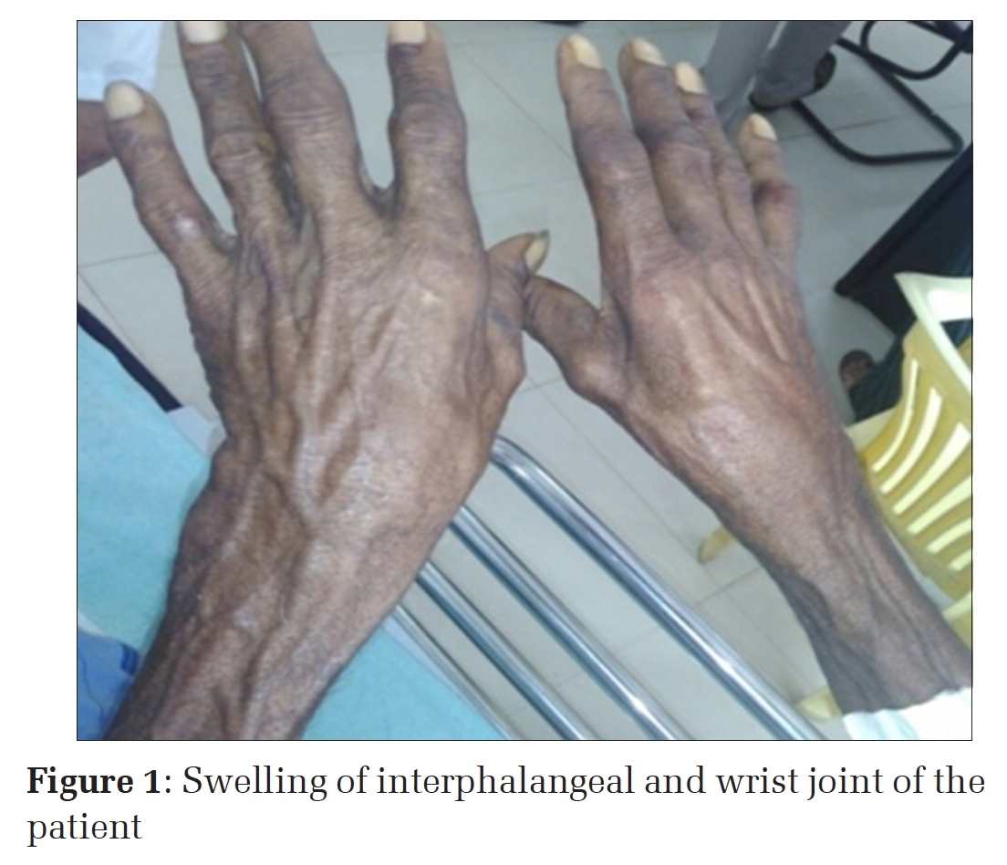
After administration of General anaesthesia, abdominal cavity was accessed by creating the pneumoperitoneum. Both the open (Hasson’s) and closed (Veress needle) method was used to create the pneumoperitoneum according to the cases. If adhesions were suspected as like the previous history of surgery, Veress needle was inserted in the Palmers point to create the pneumoperitoneum to avoid the inadvertent bowel injury. We prefer 10 mm camera port in the infra or supra umbilical region, but camera port may vary according to the suspected abdominal pathology. 2 or 3 working ports were made according to the therapeutic intervention as per laparoscopy. [Figure 1]

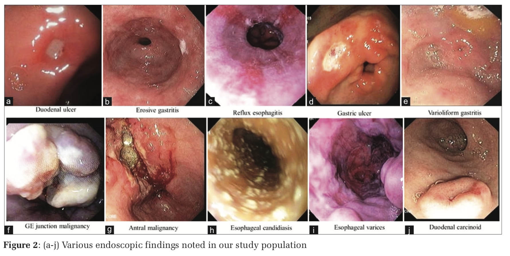
The whole abdominal cavity was carefully surveyed in a routine sequence. [Figure 2] Intra-operative findings were noted and interventions were planned according to that. Some interventions like adhesiolysis, mesenteric lymphadenectomy, biopsy, ascitic fluid analysis were done if required. In the follow up for 3 months, patients were evaluated for the clinical improvement, degree of pain relief, recurrence and complication.

All participants were evaluated for tuberculosis treatment response by assessment at week 4 and week 8 following laparoscopy. Therapeutic response was considered if there were at least two or more of the following criteria present: 1) weight gain ≥5%, 2) haemoglobin increase ≥1 gm. % and 3) at least half of the symptoms were much better or resolved, including assessment of adherence to tuberculosis treatment using the TB clinic treatment card were evaluated. All the patients had repeat abdominal ultrasonography at follow up to compare the findings with the preoperative reports.
A total of 59 patients (39 male & 20 female) who were suspected of having abdominal tuberculosis underwent diagnostic laparoscopy after obtaining informed & written consent. The highest incidence was seen in 20-29 year age group 24 (40.67%) and lowest in 50-59 year. Mean age of incidence in males was 27.45±10.19 years and 34.11±16.54 years in females with overall mean of 30.78 ±13.80 years. Males were more affected than females (male to female ratio of 1.95).
Only 7 out of 59 patients (11.86%) gave past history of pulmonary TB and 14(23.73%) patients gave positive history of presence of or treatment for pulmonary TB in other family member. None had prior history of abdominal TB.
The most common symptom was abdominal pain seen in 35 patients (59.32%) and least common was diarrhoea in 7 patients (11.86%) while pallor and abdominal tenderness were the most common signs present in 25 (42.37%) and 24 (40.67%) patients respectively with the least patients having ascitis.
X-ray chest were done in all patients but it was suggestive of tuberculosis in only 10 patients (16.94%). Most of the patients had healed pulmonary calcifications. Sputum examinations were performed only in patients with Chest X-ray suggestive of tuberculosis and all examinations were negative for tuberculosis. Standing view X-ray abdomen was done in all cases. 17(28.94%) patients showed signs of sub-acute intestinal obstruction which responded to conservative management.
The sonographic features of abdominal TB were peritoneal collection, intra-abdominal lymphadenopathy, dilated small bowel, thickened mesentery and omental thickening. Ultrasonography abdomen were done in 38 patients and all patients had evidence suggestive of abdominal tuberculosis, but histology/CBNAAT results were positive for tuberculosis in 23 patients (38.98%).
The sensitivity of ultrasonography abdomen was 92% while specificity was 61.54%. The PPV of ultrasonography abdomen was 82.14% while NPV was 80%.
In CT scan features like peritoneal free fluid, intra-abdominal lymphadenopathy, thickened small bowel loops and omental thickening were suggestive of TB. CT scan abdomen was done in all patients; 31 of them were suggestive of abdominal tuberculosis and seven were inconclusive. Only 37 of them had positive (62.71%) histology for tuberculosis and 22 were negative (37.28%).
The sensitivity and specificity of CT scan are 96% (CI 80.46-99.29%) and 46.15% (CI 23.21-70.86%) respectively. The positive predictive value and negative predictive value of CT scan was 77.42% (95% CI=60.19-88.61%) and 85.71%% (95% CI=48.69-97.43) respectively.
Mesenteric lymph nodes were biopsied from 25 patients (42.37%), 1 lymph node specimen (2.63%) from celiac group of lymph node and 1(2.63%) from peri-pancreatic group as given in Figure 3. So total 27 lymph node specimens were taken from 25 patients. Out of 27, 20 mesenteric lymph nodes had positive (74.07%) histology for tuberculosis. 7 lymph nodes (25.92%) were negative for tuberculosis. Amongst the 41 histology positive patients 29 (70.73%) had caseating granuloma and 12 had non-caseating granuloma (30%) Amongst the 18 histology negative patients, all 18 had nonspecific chronic inflammation with reactive lymph nodes.
The sensitivity and specificity of lymph nodes are 80% and 100% respectively. The positive predictive value and negative predictive value of lymph nodes are 100% (CL 83.89-100%) and 72.22% (CI 49.13-87.5%) respectively.

Small amounts of peritoneal fluid was aspirated and sent for CBNAAT from 30 patients (50.84%) and peritoneal fluid was absent in others [Figure 4]. The amount of fluid present during laparoscopy varied widely from minimal amount to 200 ml, except in two patients where it was about 500 ml and up to 20 ml of specimen was sent for tuberculosis culture from each patient.
Among 30 patients, 10 were positive (33.33%) for tuberculosis gene and 20 (66.33%) were negative. The correlation has been established between the peritoneal fluid CBNAAT and histology results. It has been found that all 10 patients had histology positive.
The sensitivity and specificity of peritoneal aspirate CBNAAT are 40% (CI=23.4-59.26%) and 100% (CI=77.19-100%) respectively. The positive predictive value and negative predictive value of peritoneal fluid CBNAAT are 100% (CI 72.25-100%) and 46.43% (CI 29.53-64.19%) respectively.

Peritoneal tubercles were present and sent for histological examination in 27 patients (45.76%). 22 of them were positive (81.48%) for tuberculosis and 5 (18.51%) were negative.
Amongst the 22 positive patients 16 had caseating granuloma (72.72%) and 6 had non-caseating granuloma (27.27%). Amongst the 5 negative patients, all had nonspecific chronic inflammation.
The sensitivity and specificity of tubercles are 56% (95%CI=37.07-73.33%) and 100% (95%CI=77.19-100%) respectively. The positive predictive value and negative predictive value of tubercles are 100% (95% CI=56-93%) and 54.17% (95% CI=28- 53%) respectively.
59 specimens (100%) were taken from omentum and sent for histological examination; 33 of them were positive (55.93%) for tuberculosis and 26 were negative (44.06%). Amongst the 33 positive patients 25 had caseating granuloma (75.75%) and 8 had non-caseating granuloma. Amongst the 26 histology negative patients, all had nonspecific chronic inflammation, one had no pathology found.
The sensitivity and specificity of omentum histology are 84% (95%CI=65.35-93.6%) and 100% (95%CI=77.19 —100%) respectively. The positive predictive value and negative predictive value of omentum are 100% (95%CI=84.15 — 100%) and 76.47% (95% CI=52.74 — 90.45%).
Total 24 patients had histology positive for tuberculosis. 1 patient had histology negative but peritoneal fluid CBNAAT positive. Total (24+1) 25 patients had positive tuberculosis findings. 13 patients were negative for tuberculosis. The differentials considered were Crohn’s disease, ulcerative colitis, carcinoma caecum, amoeboma and appendicular mass. Colonoscopy ruled out the presence of inflammatory bowel disease while CECT abdomen ruled out appendicular mass and carcinoma. The histology, AFB staining and ascitic fluid CBNAAT were all negative for tuberculosis in 13 patients, but it showed non-specific chronic inflammation in 12 and no pathology was found in one patient. Despite having no pathology in 1 case, standard empirical anti tubercular therapy was started considering the clinical findings, raised inflammatory markers and endemic nature of the disease. The histology, AFB staining and ascitic fluid CBNAAT were all positive in 2 patients. Sensitivity and specificity of these couldn’t be established because the numbers of patients are not the same in all the group of tests.
A total 5 patients (8.47%) had conversion to laparotomies for various reasons. In 3 of the cases there was huge amount of ascitic fluid which prevented proper visualisation by laparoscopy. In 1 case there was adhesions all around the bowel and omentum, so laparotomy was required for having a clearer view. In 1 case there was an accidental small bowel injury while putting the instruments which required laparotomy and subsequent suture. There was no death related to conversion to laparotomy. All 59 patients undergoing laparoscopy were started on standard anti-tuberculosis treatment without delay, whilst awaiting histology report. 48 patients were started on category 1 while 11 patients were started on category 2 treatment. Follow up of our patients was good. We followed the patient up to two months post laparoscopy. No patients died during that two months period. Out of 59 patients, only 37 patients (62.71%) came for review, all of them had positive diagnosis of tuberculosis and all of them improved symptomatically with anti TB medications. Repeat abdominal U/S, Hb%, were done and body weight were taken in all 24 patients and a trend of improvement was noted in all patients. There was no procedure related mortality during or after the laparoscopy.
It has been established that the incidence of all tuberculosis, including abdominal tuberculosis is rising and the diagnosis is still difficult.
The mortality of tuberculous peritonitis is 47- 49% if untreated, although it can be less than 5% with treatment, several studies report a mortality of up to 60% mainly because of delayed or missed diagnosis. 5, 6 To avoid any delay or missed diagnosis we wished to look at the utility of diagnostic laparoscopy in abdominal tuberculosis. Although the concept of diagnostic laparoscopy in tuberculosis is not new. 7 Historically peritoneal biopsy or peritoneoscopy. 8 Used to be done under local anaesthetics with a limited view of the abdominal cavity and higher complication rates, whereas at present laparoscopy is performed under general anaesthesia with better intra-operative views and less complication rates.
The male female ratio is highly variable from equal in the series reported by Ramesh et al to marked male predominance reported in Abdelaal A et al and to a marked female predominance in the study of Safarpor Faizollah et al. 9, 10, 11 In present study , disease incidence was found to be more in males 39(66.10%) cases, females were 20(33.90%) with male to female ratio of 1.95:1 which is comparable with study by Rai et al. 12
The most common symptom in present study was abdominal pain in 35 patients (59.32%) followed by loss of appetite and abdominal distention (51.22% each), fever (48.90%) and weight loss (45.66%) which is comparable with studies by Khan R et al, Islam J et al. 13, 14 Pallor and abdominal tenderness was common findings present in 25 (42.37%) and 24 (40.67%) patients respectively. Finding of abdominal tenderness in present study is comparable with study by Safarpor Faizollah et al. In the present study, association of old pulmonary TB with abdominal TB was found in 11.86% patients which comparable with study by Rai et al and more than in study by Ramesh J et al while family history of pulmonary TB or treatment for pulmonary TB in family member was found in 23.73% patients with abdominal TB which is comparable with study by Rai et al.
In the present study, 68.41% patients had normocytic normochromic anemia which is comparable to study by Wells AD et al 15 and Ramesh J et al while 28% patients showed raised TLC count which is comparable with Islam J et all .
The chest radiographs of only 10 patients (16.94%) were suggestive of tuberculosis with pulmonary infiltrates, though sputum cultures were negative in all 10 patients; a percentage one fourth of that reported in studies by Nafeh et al and Ramesh J et al. 8, 10 Ultrasonography of abdomen was suggestive of tuberculosis in all patients, but histology/CBNAAT results were positive in 20 patients. Though the sensitivity of CT scan was high (96%), the specificity was very low (46.15%) in our series. Caseating lymph nodes with hypodense centres and peripheral rim enhancement along with calcification are highly suggestive of tuberculosis. 16 Histology for tuberculosis was positive in 70.73% of patients with high ESR which is comparable with study by Islam J et all. 10 patients (33.33%) had positive CBNAAT from peritoneal aspirate which is comparable with study by Jin et al.
Mesenteric lymphadenopathy was the common finding in our series and 27 (71.05%) of our patients had lymph nodes biopsied which is comparable with study by Islam J et all. Tubercles were present only in 45.76% of our patients, whereas Nafeh et al had 58% and Al-Mulhim et al had 91% of their patients with tubercles. 8, 17 Adhesions were present in 12 patients (31.57%) in our series which was less than in the series by Nafeh et al (42%) and Al- Mulhim et al (52%) and more than in study by Islam J et all (22%).
The most tuberculosis positive histology specimens are tubercles (82.35%) and lymph nodes (74.07%). The most sensitive specimen is omentum (84%) and the most specific specimen is omentum (100%). The least sensitive specimen is peritoneal aspirate (40%). Thirteen patients had diagnosis other than tuberculosis (22.03%) and these 13 patients would have been started on anti-tuberculosis therapy based on clinical and radiological findings.
Our main concern is these 13 patients (22.03%) who are tuberculosis negative and had no other alternative diagnosis. 12 of them had non-specific chronic inflammation and one had no pathology. Al-Mulhim et al had 19% non-specific chronic inflammation, Nafeh et al had 3% of their patients with non-specific chronic inflammation and 7% with unsatisfactory biopsy and Islam J et al had 18% non- specific chronic inflammation.
Three patients (5.08%) had peri operative complications e.g. bleeding and difficult access and had to be converted to laparotomy. These laparotomies are for bleeding and adhesions both (7.89%). Mimica et al reported 16% conversion rate to laparotomy due to adhesions while Islam J et al reported conversion rate (11%) to laparotomy due to bleeding (33%), adhesions (22%), bowel perforation (22%) and technical difficulties (22%). 18
This study has shown the feasibility of performing laparoscopy in the majority of patients with suspected abdominal tuberculosis. It has a high yield to establish the diagnosis of abdominal tuberculosis (65.78%) by sampling macroscopically pathological tissues.
We are thankful to all the patients, OT staffs, anaesthesiologists and OT attendants for their immense help during the conduct of this study
None Declared
None
Subscribe now for latest articles and news.