Journal of Medical Sciences and Health
DOI: 10.46347/jmsh.v10.i2.23.3
Year: 2024, Volume: 10, Issue: 2, Pages: 148-153
Original Article
Rajshree Badami1 , A S Anil Kumar2 , Sundeep3
1Lead Diagnostics, Eka. Care, Regent Gateway, Seetharama Palya, Whitefield, Bangalore,
2Professor & Head, Department of Biochemistry, Kempegowda Institute of Medical Sciences, Bangalore, Karnataka, India,
3Professor & Head, Department of Opthalmology, Kempegowda Institute of Medical Sciences, Bangalore, Karnataka, India
Address for correspondence:
Rajshree Badami, Lead Diagnostics, Eka. Care, Regent Gateway, Seetharama Palya, Whitefield, Bangalore.
E-mail: [email protected]
Received Date:01 December 2023, Accepted Date:18 May 2024, Published Date:17 June 2024
Aim: The study aims to determine whether oxidative stress contributes significantly to the development of Senile cataract by comparing serum levels of oxidative stress marker Malondialdehyde and antioxidants Ascorbic acid, Alpha Tocopherol and Uric acid among Senile cataract patients and healthy controls. Materials and Methods: A total of 86 subjects were included in the study, out of which 49 were senile cataract patients and 37 were age and gender matched healthy controls. Estimation of Malondialdehyde, Ascorbic acid, Alpha- Tocopherol and Uric acid was done in the serum samples of all subjects included in the study. Results: Mean MDA level in cases was significantly higher at 4.41nmol/ml compared to controls which was 3.88nmol/ml. Mean ascorbic acid level among cases was 10.99 mg/L whereas in controls it was significantly higher at 13.69 mg/L. α-tocopherol level among cases was 7.98 mg/L whereas in controls it was significantly higher at 8.76 mg/L. Mean uric acid level among cases was 4.29mg/dl whereas in controls it was 4.55mg/dl, the difference was statistically insignificant. Conclusion: Oxidative stress and antioxidant status contributes significantly to the development and progression of senile cataract. Clinical Significance: Measures to monitor oxidative stress and improve antioxidant status in the population can be helpful in delaying the onset and progression of senile cataract.
Keywords: Oxidative stress, Senile cataract, Malondialdehyde, Antioxidants
Cataract is defined as “a complete or partial opacification on or in the lens or its capsule.” It is the worldwide leading cause of reversible visual impairment, accounting for 50% of population suffering from blindness 1, 2, 3.
In developing countries, 50-90% of all blindness is due to cataract 2. In tropical countries, cataracts evolve earlier in life and are three times more prevalent than other countries. In India, the estimated annual incidence of cataract is 4 million. Out of these, only 2.5 million get operated leaving about 1.5 million untreated 3, 4.
Senile or age-related cataract is the most common type of cataract 5, 6. Currently there is no medical treatment for senile cataract and surgery remains the only choice of treatment 1, 2 . Prevention or a delay of 10 years in the development of senile cataract will reduce its prevalence by 50% thereby improving quality of life and reducing economic burden due to disability and surgery 3, 4.
Oxidative stress is often speculated to be one of the initiating factors in the development of age-related cataracts. Some most important oxidants are free radicals. When the harmful effects produced due to oxidants exceed the body’s capacity to eliminate or scavenge them, it leads to oxidative stress 1, 5, 7.
The lens is constantly exposed to light and oxygen and is at a high risk for photo oxidative damage. This damage increases with the ageing lens 1, 8. The major effect of oxidative stress on the lens includes damage to the membranes of the lens epithelium by peroxidation of membrane lipids. Oxidative damage to lens fiber proteins causes protein aggregation resulting in loss of transparency 1, 6, 8.
Lipid peroxidation represents oxidative damage to membrane lipids including membranes of lens fibers 9. Malondialdehyde (MDA) is an important stable end-product of free radical reaction on the membrane fatty acids and can be used as a reliable marker of lipid peroxidation 7.
The lens antioxidant defense system includes non-Enzymatic as well as Enzymatic antioxidants. Vitamin C (Ascorbic acid), Vitamin E (α-tocopherol) are the nutritional non-Enzymatic antioxidants whereas Uric acid and reduced Glutathione are important endogenous non-Enzymatic antioxidants. Superoxide Dismutase (SOD), Catalase and Peroxidases are the important endogenous Enzymatic antioxidants 1, 2, 3, 8, 10, 11.
Ascorbic acid is an important antioxidant in extracellular fluids. It prevents lipid peroxidation by its action against aqueous peroxyl radical. Ascorbic acid helps in regenerating α-tocopherol by converting α- tocopheroxyl radical produced during free radical scavenging, back to α-tocopherol 6.
α-Tocopherol is a major lipid soluble antioxidant present in the cell membranes that protects them against lipid peroxidation. α-tocopherol is a chain breaking antioxidant that directly scavenges free radicals 6.
Uric acid is the end -product of purine nucleotide metabolism. Uric acid plays the antioxidant role by scavenging singlet oxygen species and its antioxidant effect is being recognized in senile cataract. However, the overproduction of uric acid itself can cause oxidative stress as the pathway that forms uric acid also generates hydrogen peroxide 11.
One of the aims of this study, hence, is to look for evidence of ongoing lipid peroxidation in senile cataract by estimating and comparing serum MDA levels among subjects of senile cataract with age and gender matched healthy controls.
Further, this study aims to determine if the serum levels of nutritional antioxidants ascorbic acid, α-tocopherol differ significantly between cataract subjects as compared to healthy controls. Lastly, the study aims to look for correlation, if any, between the levels of MDA and the antioxidants in cataract subjects as well as in healthy controls.
Ethical clearance was obtained by the Institutional Ethical committee. A case control study was carried out in the Department of Ophthalmology, Kempegowda Institute of Medical Sciences and Research Centre, Bangalore from February 2014 to September 2015.
Informed consent was obtained from all the participants prior to their inclusion in the study. All subjects underwent a general physical and ophthalmologic examination before being chosen to participate in the study. Subjects in the age group of 50-80 years, with newly diagnosed senile cataract were included in the study. Healthy, age and gender matched volunteers served as controls for the study.
A total of 86 subjects were included in the study out which 49 subjects were diagnosed with senile non-pathologic cataract in the age group of 50-80 years. The control group comprised of 37 age and gender matched healthy volunteers.
Subjects with known genetic predispositions to cataract were excluded. All subjects came from an agricultural background with comparable UV exposure levels.Cases of secondary cataract due to diabetes, metabolic disorders, radiation, glaucoma, trauma and steroid therapy were excluded from the study. Subjects with concomitant ocular infections, allergies, inflammation, contact lens users, history of ocular surgery and any posterior segment pathologies were also excluded. Subjects who reported dietary supplement use within last 6 months, particularly antioxidants, were excluded from the study to eliminate confounding effects on oxidative stress. Subjects presenting with history and general physical examination suggestive of hypertension, endocrine disorders, hepatic, gastrointestinal or renal dysfunction, gout, autoimmune diseases and malignancy were excluded.
Fasting blood and urine samples were collected from all subjects for estimation of glucose to screen them for diabetes; subjects with normal fasting blood glucose levels were included in the study. Subjects with a positive urine sugar or urine protein result on dipstick examination were also excluded.
Subjects consuming alcohol, smokers, those on exogenous hormones, multi-vitamin medication, antioxidant supplements and systemic uric acid-lowering therapy were also excluded from the study.
Sample collection: Following an overnight fast, 10 ml of venous blood was collected by venipuncture, under septic precautions, using a disposable syringe and needle. Serum was separated by centrifugation at 3500 revolutions per minute (rpm) for 10 minutes at room temperature. Samples were stored at -20oC until analysis.
Analysis: Serum malondialdehyde was estimated using Thiobarbituric Acid Method 12, 13, 14, 15, 16 . Ascorbic acid was estimated using 2, 4- Dinitrophenyl hydrazine method 17, 18, 19 . Serum α-tocopherol was estimated by Baker and Frank Method 20, 21, 22. Serum Uric acid was estimated by Enzymatic colorimetric method using Uricase 23.
Statistical Analysis: The data collected was analyzed statistically using IBM SPSS v22 statistical software. Statistics namely mean, standard deviation, range were computed. Any significant difference between the mean values of the study group and the control group was tested using independent sample student t-test. Correlation between MDA and antioxidants was tested using Spearman’s rank correlation test. A p value of < 0.05 was considered statistically significant.
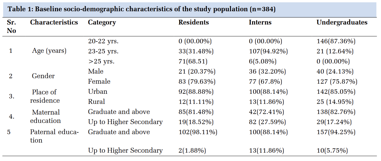
Among the cases, 27 (55%) were male and 22 (45%) were female. The control group comprised of 20 (54%) male and 17 (46%) female subjects.
The mean age group of cases was 64.06 years while the mean age group of controls was 61.78 years. Mean age of male and female cataract subjects was 63.4 years and 64.9 years respectively. The controls had a mean age of 62.6 years among males and 60.8 years among females.
|
|
Group |
|
Mean (SD) |
Std. Deviation |
t |
df |
p-value |
|
MDA (nmol/ml) |
Case |
49 |
4.41 (0.92) |
0.52 (0.14, 0.91) |
2.728 |
84 |
0.008* |
|
Control |
37 |
3.88 (0.83) |
|
|
Group |
|
Mean (SD) |
Std. Deviation |
t |
df |
p-value |
|
Vit C (mg/L) |
Case |
49 |
10.99 (3.03) |
-2.69 (-4.05, -1.33) |
-3.934 |
84 |
<0.001* |
|
Control |
37 |
13.69 (3.28) |
|||||
|
Vit E (mg/L) |
Case |
49 |
7.98 (1.58) |
-0.77 (-1.46, -0.08) |
-2.226 |
84 |
0.03* |
|
Control |
37 |
8.76 (1.62) |
|||||
|
Uric Acid (mg/dl) |
Case |
49 |
4.29 (1.11) |
-0.25 (-0.74, 0.22) |
-1.061 |
84 |
0.29(NS) |
|
Control |
37 |
4.55 (1.12) |
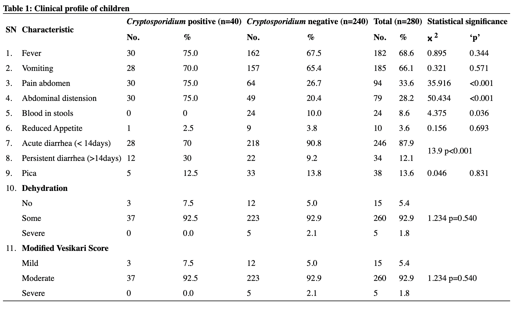
MDA levels: The mean MDA level in cases was 4.41nmol/ml compared to controls which was 3.88nmol/ml. The difference was statistically significant.
Ascorbic acid levels: Mean ascorbic acid level among cases was 10.99 mg/L whereas in controls it was significantly higher at 13.69 mg/L.
α-Tocopherol levels: α-tocopherol level among cases was 7.98 mg/L whereas in controls it was significantly higher at 8.76 mg/L.
Uric acid levels: Mean uric acid level among cases was 4.29mg/dl whereas in controls it was 4.55mg/dl. The results were statistically not significant.
Among cases, no correlation was found between MDA and any of the antioxidants. Among controls, a significant positive correlation was obtained between MDA and α-tocopherol levels.
Detection of oxidative stress can be done by quantifying byproducts of free radical injury. MDA is one such marker that provides an indirect evidence of cell membrane damage due to lipid peroxidation.
Our study revealed that mean serum MDA levels were significantly higher in cataract subjects (4.41 + 0.92 nmol/ml) when compared to healthy controls (3.88 + 0.83 nmol/ml), indicating increased ongoing lipid peroxidation in cataract subjects. Few other studies recorded similar results 1, 2, 3, 5, 6, 7, 8, 9, 10, 24, 25, 26.
Oxidative stress is a result of the imbalance between mechanisms that generate free radicals and those that scavenge them. Antioxidants combat free radicals and play a protective role against free radical injury 27.
In our study we estimated two exogenous antioxidants – ascorbic acid and α-tocopherol and one endogenous antioxidant – uric acid.
Ascorbic acid is a powerful aqueous phase antioxidant that efficiently scavenges peroxyl and superoxide radicals with a slightly weaker activity against hydroxyl radicals. Ascorbic acid also regenerates other antioxidants - glutathione and α-tocopherol that play an important antioxidant role in the lens 28.
Our study revealed significantly lower mean serum ascorbic acid level in cataract subjects (10.99 + 3.03 mg/L) compared to controls (13.69 + 3.28 mg/L). Similar results were obtained by Pradhan et al, Kawanpure et al and Selvi et al, whereas the study by Garg et al concluded a non-significant reduction in vitamin C levels 2, 5, 6, 9, 10.
α-tocopherol is the principle exogenous antioxidant in the lipid phase and a powerful peroxyl scavenger and chain breaking antioxidant, most effective in preventing lipid peroxidation 29.
We obtained significantly lower mean α-tocopherol levels in cataract subjects (7.98 + 1.58 mg/L) as compared to controls (8.76 + 1.62 mg/L) in our study. Similar results were recorded by Selvi et al, Kawanpure et al and Katta et al. However, studies by Chang et al, Garg et al, Pradhan et al, Gale et al, Olmedilla et al and Knekt et al recorded statistically insignificant results 2, 3, 5, 6, 9, 10, 30, 31, 32.
Uric acid is the end-product of purine metabolism. It is an endogenous antioxidant, acting as a free radical scavenger and transition metal chelator in the extracellular environment. Paradoxically however, elevated intracellular levels of uric acid have a pro-oxidant effect 33.
The difference in the mean uric acid levels among cataract subjects (4.29 + 1.11mg/dl) and controls (4.55 + 1.12mg/dl) in our study was statistically insignificant. Adedapo et al obtained significantly reduced uric acid levels in cataract subjects as compared to controls 4.
Our study did not reveal a significant correlation between the levels of MDA and antioxidant parameters among cataract subjects. However, a positive correlation was obtained between MDA and α-tocopherol levels among controls. Higher α-tocopherol levels may be a cause as to why the control subjects do not show signs and symptoms of cataract, even in the presence of higher MDA levels. This suggests a possible delay in the appearance of the symptoms of cataract, among subjects with higher α-tocopherol levels 9, 34.
Significant association between antioxidant supplementation and prevalence of age related cataract has been documented, suggesting a role of dietary factors in the development and prevention of senile cataract 35, 36, 37.
Valero et al studied the intake of dietary antioxidants among cataract subjects and healthy controls and concluded that increased dietary antioxidant intake was significantly associated with lower prevalence of cataract 35.
Taylor et al obtained inverse association between dietary and supplemental antioxidant intake with prevalence of cortical opacities in women less than 60 years of age 36.
Robertson et al demonstrated that cataract subjects were consuming significantly less amounts of supplementary antioxidants compared to healthy controls 37.
Goyal et al studied the direct effect of vitamin C and vitamin E on the lens epithelium obtained from surgically removed cataractous lenses and demonstrated a reduction in catalase activity in the lens epithelium when exposed to vitamin C and E 38.
Subscribe now for latest articles and news.