Journal of Medical Sciences and Health
DOI: 10.46347/jmsh.2020.v06i02.008
Year: 2020, Volume: 6, Issue: 2, Pages: 43-45
Original Article
Archana Narasipuram1, Mrudula Chandraputula2, Hajira Fatima3
1Assistant Professor, Department of Anatomy, Apollo Medical College, Hyderabad, Telangana, India,
2Professor and Head, Department of Anatomy, Apollo Medical College, Hyderabad, Telangana, India,
3Tutor, Department of Anatomy, Apollo Medical College, Hyderabad, Telangana, India
Address for correspondence:
Archana Narasipuram, Plot No: 181/1, The Pride, 2nd floor, O.U Colony, Shaikpet, Hyderabad - 500 008, Telangana, India. E-mail: [email protected]
Introduction: Pulmonary veins originate from the alveolar capillary plexus and carry oxygenated blood to the left atrium. The fibrous pericardium is perforated by the pulmonary veins and they drain separately in the posterosuperior aspect of the left atrium as the right and left pulmonary veins. From the dorsal atrial wall, primordial vein arises as an outgrowth just to the left of the septum primum. The primordial pulmonary vein and its main branches are incorporated into the wall of the left atrium as the atrium expands and results in the formation of four pulmonary veins. The knowledge of the variations in the pulmonary veins is helpful for endoscopists and also for the surgeons operating for arrhythmias. The aim of the study was to assess the number of pulmonary veins opening into the left atrium and their variations.
Materials and Methods:The present study was conducted in the Department of Anatomy, Apollo Medical College, Hyderabad. Study design: This study is an observational study; the study material comprised 30 embalmed cadaveric heart specimens of unknown sex. Old and damaged cadaveric heart specimens were excluded from the study. The number of pulmonary veins opening into the left atrium and their variations was observed. The percentage of variations on the right and left side was calculated.
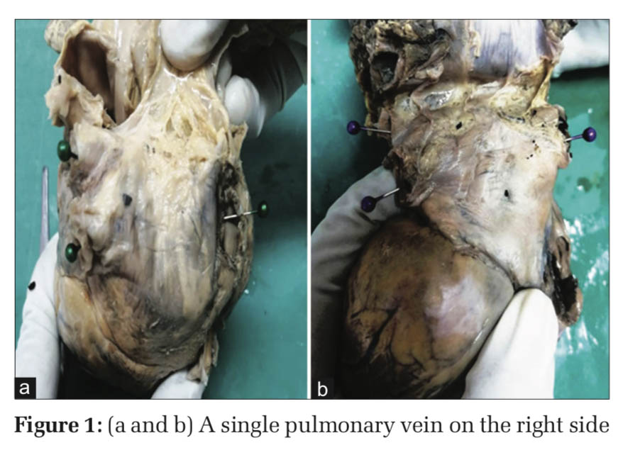
Results: In the present study, out of 30 heart specimens which were used for the study, four pulmonary veins draining into the left atrium were observed in 26 heart specimens which are a normal pattern. A variation in the number of pulmonary veins opening into the left atrium was observed in only four heart specimens which were of a different pattern. The variation in the number of pulmonary veins in all the four heart specimens was on the right side.
Conclusion: Knowledge about the variations in number of pulmonary veins is helpful as pulmonary veins are an important source of ectopic atrial electrical activity and can be useful for cardiologists, radiologists, and cardiothoracic surgeons in radiofrequency ablation surgeries.
KEY WORDS:pulmonary veins, ectopic electric activity, ablation surgeries
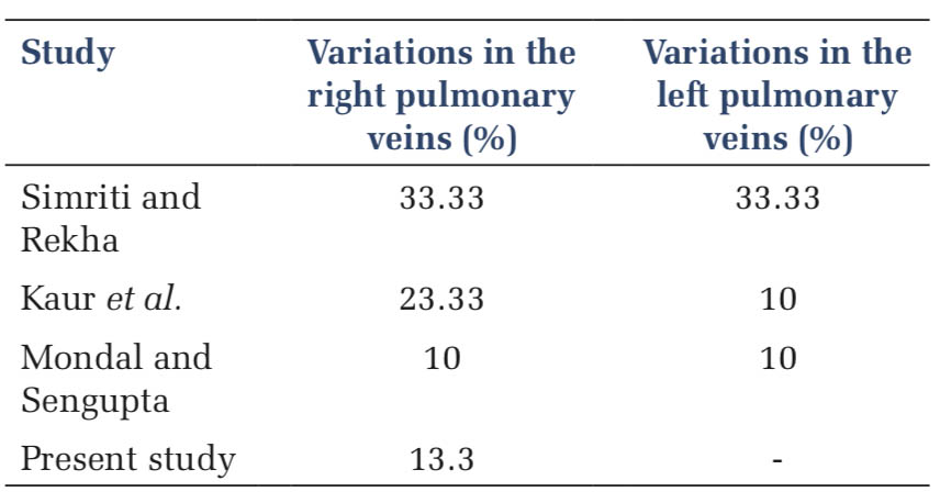
IntroductionPulmonary veins originate from the alveolar capillary plexus and carry oxygenated blood to the left atrium. The fibrous pericardium is perforated by the pulmonary veins and they drain separately in the posterosuperior aspect of the left atrium as the right and left pulmonary veins.[1] The number of pulmonary veins may differ due to variable incorporation of the primitive pulmonary vein into the left atrium.[1] The terminal parts of the pulmonary veins are surrounded by atrial myocardium; these areas represent potential accessory reentrant circuits responsible for the initiation or maintenance of supraventricular tachycardia or atrial fibrillation and may be percutaneously ablated.[1] The smoothness of the left atrial wall is attributed to the primordial vein incorporation into the left atrium. From the dorsal atrial wall, primordial vein arises as an outgrowth just to the left of the septum primum.[2] The primordial pulmonary vein and its main branches are incorporated into the wall of the left atrium as the atrium expands and results in the formation of four pulmonary veins.[2] Previously, the variations in the number of the pulmonary veins were considered as rare and they were reported in few case reports.[3] Variations in pulmonary venous anatomy were noticed in 36% of the patients in recent studies.[4] The knowledge of the variations in the pulmonary veins is helpful for endoscopists and also for the surgeons operating for arrhythmias. The present study was done to assess the number of pulmonary veins opening into the left atrium and their variations. Materials and MethodsThe present study was conducted in the Department of Anatomy, Apollo Medical College, Hyderabad. ResultsIn the present study, out of 30 heart specimens which were used for the study, four pulmonary veins draining into the left atrium were observed in 26 heart specimens which are a normal pattern. A variation in the number of pulmonary veins opening into the left atrium was observed in only four heart specimens which were of a different pattern. The variation in the number of pulmonary veins in all the four heart specimens was on the right side. There is only one pulmonary vein on the right side of the left atrium in three specimens [Figure 1a and b]. In one specimen, it appeared as if the two pulmonary veins fused to form a single opening [Figure 2]. One specimen showed four openings on the right side and two openings on the left side [Figure 3]. DiscussionThe pulmonary veins are responsible for carrying oxygenated blood from the lungs to the left atrium of the heart. The pulmonary veins differ from the other veins in the body that usually carries deoxygenated blood from rest of the body to the heart. Humans have four pulmonary veins, right pulmonary veins as right superior and right inferior and left superior, and left inferior veins. Initially, the dorsal wall of the left atrium receives a single large pulmonary vein.[5] The right and left pulmonary veins are the branches from the main pulmonary vein and each branch further divides into upper and lower branches. Hence, dorsal wall of the left atrium is formed by the incorporation of four pulmonary veins into the atrium.[5] Incorporation of the pulmonary veins beyond their first division results in the development of supernumerary or accessory pulmonary veins and such variations are usually found on the right side.[6] In the present study, 10% of specimens showed a single pulmonary vein on the right side and 3% of the specimens showed four pulmonary veins on the right side. Similarly, in a cadaveric study conducted by Parsana et al., single pulmonary vein on the right side was observed in 14% of the specimens and four pulmonary veins in 4%. In a study conducted by Kaur et al., three pulmonary veins were found in 13.3% specimens and four pulmonary veins in 6.67% and 5 pulmonary veins in 3.3%. In a study conducted by Simriti and Rekha,[7] single pattern of pulmonary veins was seen in 20% hearts, double pattern observed in 66.6% hearts, and triple pattern was seen in 13.3% hearts on the right side. About 10% showed variations in the right in a study by Mondal and Sengupta.[8] Most of the previous studies are similar to the present study.
|
ConclusionKnowledge about the variations in number of pulmonary veins is helpful as pulmonary veins are an important source of ectopic atrial electrical activity and can be useful for cardiologists, radiologists, and cardiothoracic surgeons in radiofrequency ablation surgeries. |
Subscribe now for latest articles and news.