

Journal of Medical Sciences and Health
DOI: 10.46347/jmsh.2018.v04i03.005
Year: 2018, Volume: 4, Issue: 3, Pages: 26-28
Case Report
K Suman, Namrata Rao
Department of Pathology, Melaka Manipal Medical College, Manipal Academy of Higher Education, Manipal, Karnataka, India
Address for correspondence:
K Suman, Department of Pathology, Melaka Manipal Medical College, Manipal Academy of Higher Education, Manipal-576104
Renal oncocytoma is a benign tumor which arises from collecting duct cells. It may be detected incidentally or present with hematuria and flank pain. It is a rare occurrence in pregnancy. Here, we present a case report of a 26-year-old patient who was detected with a renal mass in pregnancy and radical nephrectomy was performed 5 months after delivery. Gross examination of the kidney showed a well-circumscribed tan brown tumor. Microscopy revealed neoplastic oncocytes arranged in nests, alveolar, and tubular pattern having round central nuclei with evenly dispersed chromatin and abundant eosinophilic granular cytoplasm. On immunohistochemistry, these cells were positive for CD117 and negative for CK7, CD10, S100, and Hale’s colloidal iron stain. Diagnosis of oncocytoma was given.
KEY WORDS:Incidental, oncocytoma, renal mass.
Renal oncocytoma is an unusual benign tumor first described by Zippel in 1942. They are usually solitary masses and represent between 3% and 10% of all renal tumors. Clinically, oncocytoma may be asymptomatic. A few patients may present with hematuria, flank pain, or palpable mass.[1] It is composed of oncocytes that exhibit a granular eosinophilic cytoplasm.[2] Diagnosis of cancer during pregnancy is rare with cervical cancer and breast cancer being the most commonly identified cancers. Among urological tumors, which are rarely identified during pregnancy, renal cell carcinoma is the most common tumor followed by angiomyolipoma.[3,4] Only very few cases of oncocytoma diagnosed during the antenatal period have been reported; hence, we report this case for its rarity.
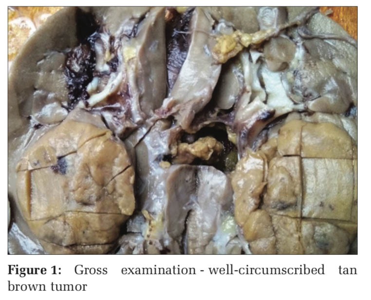
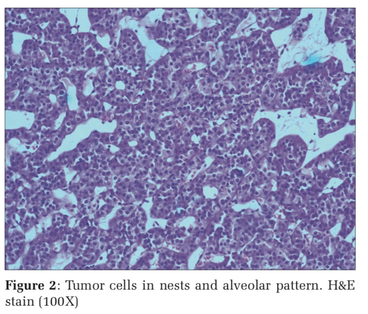
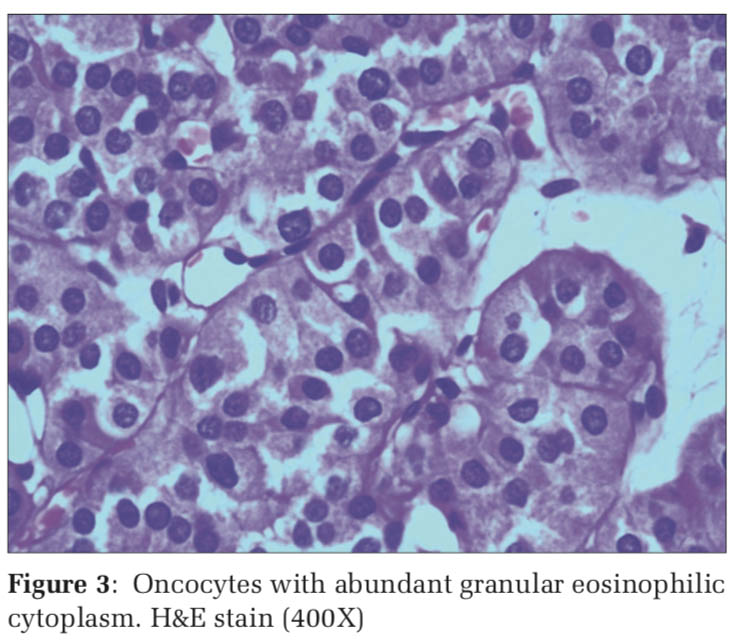
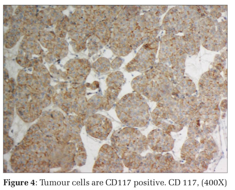
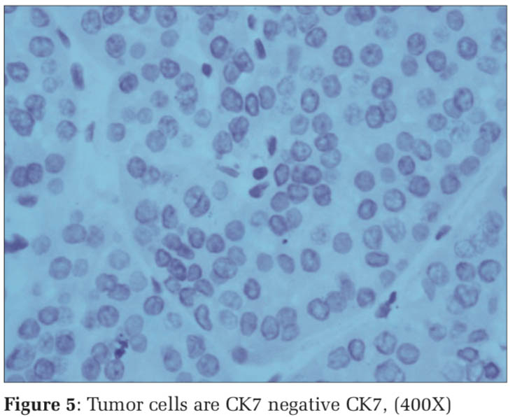
A 26-year-old female patient was incidentally detected with the left renal mass on ultrasonography during routine antenatal examination. 5-month post-delivery by lower segment cesarean section, the patient was evaluated by contrast computed tomography of abdomen and pelvis which showed a non-homogenously enhancing isodense mass lesion in the left kidney. The patient underwent laparoscopic radical nephrectomy. On gross examination, the cut section of kidney showed a unifocal, well- circumscribed tan brown tumor measuring 5.5 cm × 4.5 cm × 4 cm with focal hemorrhagic areas [Figure 1]. The tumor along with adjacent parenchyma, renal sinus, perinephric fat, ureter, and renal vessels was adequately sampled. Microscopy revealed neoplastic polygonal cells arranged in nests, alveolar, and tubular pattern having round central nuclei with mild anisonucleosis, evenly dispersed chromatin and abundant eosinophilic granular cytoplasm [Figures 2 and 3]. Mitosis was rare and necrosis was absent. On immunohistochemistry, the tumor cells were positive for CD117 and negative for CK7, CD10, S100, and Hale’s colloidal iron stain [Figures 4 and 5]. Perinephric fat, renal sinus, ureter, and vessels were free of tumor. Diagnosis of oncocytoma was made based on the above findings.
Most of renal oncocytomas are asymptomatic at presentation and are discovered incidentally hematuria and pain occur in a minority of patients. Renal oncocytomas usually appear as solitary lesions measuring 4–8 cm. Multifocal and bilateral appearance have been observed in 4–6% and 4%, respectively.[1] Even when very large, they are generally well encapsulated and are rarely invasive or associated with metastases.[5] If confronted with a renal tumor during pregnancy, multidisciplinary team must be taken into account when establishing a management plan. The influence of pregnancy hormones on oncocytomas is not clear although the growth of angiomyolipomas in pregnancy has been described.[4]
Radical or partial nephrectomy is performed on the majority of patients, based on their clinical circumstances.[2]
Oncocytomas appear to originate from collecting duct cells. In most cases, oncocytomas and different histological subtypes of renal cell carcinoma can be differentiated on gross inspection and microscopy. Sometimes, the differentiation is difficult, especially among the eosinophilic and granular variants of conventional renal cell carcinoma and oncocytoma. Chromophobe renal cell carcinoma shows strong diffuse positivity to Hale’s colloidal iron stain, whereas oncocytoma shows negative or weak focal staining. Oncocytomas are typically CK7 negative and CD117 positive, while most chromophobe renal cell carcinoma are positive for CK7 and CD117 negative.
Renal oncocytoma is a benign, generally unifocal tumor arising from the collecting duct cells and is usually treated by partial or radical nephrectomy.
Subscribe now for latest articles and news.