Journal of Medical Sciences and Health
DOI: 10.46347/jmsh.v12.i1.25.230
Year: 2026, Volume: 12, Issue: 1, Pages: 51-54
Original Article
Shrish Bhatnagar 1, Aashlesha Kritika 1, Sumaiya Shamsi 1, Sachin Khanduri 2
1Department of Pediatrics, Eras Lucknow Medical College and Hospital, Lucknow, UP, India.
2Department of Radio diagnosis, Eras Lucknow Medical College and Hospital, Uttar Pradesh, India.
Address for correspondence:
Shrish Bhatnagar, Department of Pediatrics, Eras Lucknow Medical College and Hospital, Lucknow, UP, India.
E-mail: [email protected]
Received Date:23 June 2025, Accepted Date:16 January 2026, Published Date:30 March 2026
Background: Accurate assessment of liver and spleen size is crucial for diagnosing hepatosplenomegaly in newborns. While ultrasonography (USG) is the gold standard, clinical methods (palpation-percussion) remain widely used in resource-limited settings. Objectives: To compare liver span measurements by palpation-percussion versus USG, establish normative data, and correlate liver/spleen size with anthropometric parameters in healthy term newborns. Methods: A cross-sectional study of 434 newborns (gestational age: 37–41 weeks) was conducted. Liver span was measured via palpation-percussion and USG; spleen size via USG. Anthropometric parameters were recorded. Statistical analysis included paired t-tests and Pearson correlation. Results: Mean liver span by palpation-percussion (5.2 ± 0.14 cm) was significantly lower than by USG (5.42 ± 0.22 cm; p < 0.0001). Spleen size averaged 4.09 ± 0.18 cm. No significant correlations existed between liver/spleen measurements and anthropometric parameters. Percentile charts were established. Conclusion: Clinical methods underestimate liver span but remain reliable for initial screening. Normative data aid in detecting hepatosplenomegaly in term newborns.
Keywords: Liver span, Spleen size, Newborn, Ultrasonography, Palpation-percussion
The liver, the largest internal organ, performs over 500 vital functions, including metabolism, detoxification, and immunity. Its size reflects physiological status and pathological conditions like congenital infections, metabolic disorders, or heart failure. Accurate assessment of liver and spleen size is crucial for diagnosing hepatosplenomegaly in newborns. While ultrasonography (USG) is the gold standard, clinical methods (palpation-percussion) remain widely used in resource-limited settings[1, 2, 3]. In newborns, hepatomegaly may indicate critical illnesses (e.g., biliary atresia, glycogen storage disease)[4, 5]. Clinical evaluation via palpation-percussion is routine but prone to inter-observer variability and inaccuracy[6]. Ultrasonography offers precision but is less accessible in resource-limited settings[7, 8].
Existing studies report conflicting correlations between liver/spleen size and anthropometric parameters (e.g., age, weight, height)[9, 10]. Most data derive from older children, creating a gap in normative standards for newborns. This study aimed to:
1. Compare liver span measurements by palpation-percussion versus USG.
2. Establish normative percentiles for liver/spleen size in term newborns.
3. Correlate measurements with anthropometric parameters (weight, length, gender, BSA).
A cross-sectional study was conducted in the Department of Pediatrics and Neonatology at Era's Lucknow Medical College and Hospital between October 2019 and December 2021. Ethical approval was obtained from the Institutional Ethical Committee, and written informed consent was secured from parents “ECR/717/Inst./UP/2015/RR-21) R Cell/EC 2020/41 dated 18.04.2020 ”. The study included 434 healthy term newborns (gestational age: 37–41 weeks) with birth weights >2.5 kg. Exclusion criteria encompassed congenital anomalies, critical illness, or abdominal defects identified via ultrasonography. The sample size was calculated using standard deviations from prior
studies[3] to achieve 90% power at α = 0.05.
Anthropometric parameters recorded were weight (kg), length (cm), head circumference (cm), chest circumference (cm), mid-upper arm circumference (MUAC, cm), body surface area (BSA, calculated via Mosteller formula[11]), and transcutaneous bilirubin (TcB, g/dL). Liver span was assessed using two methods: (1) clinical palpation-percussion along the right midclavicular line (MCL) during inspiration, where the upper and lower borders were marked by dullness-to-tympany transitions and measured with a flexible tape; and (2) ultrasonography (USG) using a Siemens LOGIQ P9 (3–5 MHz probe), with longitudinal dimensions in the MCL (mean of three measurements). Spleen size was measured via USG in the coronal view.
Statistical analyses were performed using SPSS v25.0. Continuous variables were expressed as mean ± SD and median (IQR). Paired t -tests compared liver span measurements between methods, while Pearson correlation assessed relationships between liver/spleen size and anthropometric parameters. Percentiles (3rd–97th) were computed for all measurements, with significance set at p < 0.05.
The cohort comprised 434 newborns: 224 males (51.6%) and 210 females (48.4%). Most infants (78.8%) were assessed on day 2 of life. Mean gestational age was 38.4 ± 1.2 weeks. Anthropometric data are summarized in [Table. 1].
Liver span measured by palpation-percussion (mean: 5.2 ± 0.14 cm; range: 4.97–5.63 cm) was significantly lower than USG measurements (mean: 5.42 ± 0.22 cm; range: 5.07–5.8 cm; p < 0.0001; mean difference: −0.23 cm; 95% CI: −0.25 to −0.20). Percentile values for both methods are detailed in [Table. 3]. Spleen size averaged 4.09 ± 0.18 cm (range: 3.77–4.43 cm), with no gender-based differences observed (percentiles in [Table. 4]).
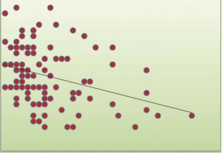
No significant correlations existed between liver span (clinical or USG) and anthropometric parameters (birth weight, length, BSA, head circumference; r = 0.007–0.079; all p > 0.05; [Table. 5]). Similarly, spleen size showed no correlations with anthropometry (r = −0.028–0.077; all p > 0.05).
| Parameter | Mean ± SD | Median (IQR) | Range |
|---|---|---|---|
| Birth weight (kg) | 3.04 ± 0.29 | 3.08 (2.8–3.3) | 2.6–3.67 |
| Length (cm) | 49.01 ± 1.49 | 48.5 (48–50) | 47–52 |
| BSA (m²) | 0.33 ± 0.04 | 0.33 (0.29–0.36) | 0.27–0.38 |
| Head circumference (cm) | 34.05 ± 1.06 | 34.5 (33–35) | 32–35.5 |
| Chest circumference (cm)| | 32.03 ± 1.04| | 32 (31–33.5) | 31–34 |
| MUAC (cm) | 8.97 ± 0.17 | 9 (8.8–9.2) | 8.7–9.2 |
| TcB (g/dL) | 0.82 ± 0.53 | 1 (0.4–1) | 0–3 |
| Liver span(cm) | Mean Difference | Std. Deviation | Std. Error Mean |
95% Confidence Interval of the Difference |
t value | p value | |
|---|---|---|---|---|---|---|---|
| Lower | Upper | ||||||
| Liver Span (palpation, percussion method)-Liver Span (ultrasonography method) | -0.2265 | 0.25347 | 0.01217 | -0.2504 | -0.2026 | -18.616 | <0.0001* |
| Liver Span | N | Mean | Std. Deviation | Minimum | Maximum |
Percentiles |
|||||||
|---|---|---|---|---|---|---|---|---|---|---|---|---|---|
| 3 | 5 | 10 | 50 | 75 | 90 | 95 | 97 | ||||||
| Palpation, percussion method | 434 | 5.2 | 0.14 | 4.97 | 5.63 | 5.07 | 5.07 | 5.07 | 5.17 | 5.23 | 5.29 | 5.63 | 5.63 |
| Ultrasonography method | 434 | 5.42 | 0.22 | 5.07 | 5.8 | 5.10 | 5.10 | 5.13 | 5.37 | 5.63 | 5.70 | 5.77 | 5.77 |
| Variable | Mean ± SD | Median(25th-75th percentile) | Range |
|---|---|---|---|
| Spleen size(cm) | 4.09 ± 0.18 | 4.07(3.967-4.233) | 3.77-4.43 |
| Percentiles | 10th 3.80–3.84 cm | 50th 4.07–4.13 cm | 90th 4.33–4.40 cm |
| Variables | Birth weight (kg) | Length (cm) | BSA (m²) | Head circumference (cm) | Chest circumference (cm) | MUAC (cm) | TcB (g/dL) |
|---|---|---|---|---|---|---|---|
| Liver span in cm (palpation, percussion method) | |||||||
| Correlation coefficient | 0.073 | 0.043 | 0.079 | 0.071 | 0.032 | 0.069 | 0.008 |
| P value | 0.131 | 0.367 | 0.101 | 0.138 | 0.503 | 0.149 | 0.872 |
| Liver span in cm (ultrasonography method) | |||||||
| Correlation coefficient | 0.031 | 0.035 | 0.034 | 0.025 | 0.035 | 0.031 | 0.007 |
| P value | 0.520 | 0.464 | 0.486 | 0.608 | 0.471 | 0.521 | 0.889 |
Pearson correlation coefficient
This cross-sectional study provides robust normative data for liver span and spleen size in healthy term newborns, addressing a significant gap in neonatal assessment literature. The key finding that palpation-percussion consistently underestimates liver span by 0.23 cm compared to ultrasonography (5.2 cm vs. 5.42 cm; p < 0.0001) aligns with prior studies by Joshi et al.[1] and Amatya et al.[2]. This discrepancy stems from inherent limitations of clinical methods, including operator dependency in identifying dullness-to-tympany transitions and technical challenges in newborns with rapid respiratory patterns. Despite this systematic bias, the strong correlation (r = 0.88) between clinical and ultrasonographic measurements reported by Amatya et al.[2] supports the continued utility of palpation-percussion as a pragmatic frontline screening tool, particularly in resource-limited settings where immediate ultrasonography access is unavailable.
The mean spleen length of 4.09 cm corroborates reference ranges established by Kahramaner et al.[3], demonstrating consistency across geographically diverse neonatal populations. Notably, the absence of significant correlations between liver/spleen dimensions and anthropometric parameters (e.g., birth weight r = 0.031, BSA r = 0.034) contrasts with studies in older children[4-6]. This divergence likely reflects the physiological homogeneity of term newborns, where narrow anthropometric ranges constrain detectable associations, suggesting organ growth may follow intrinsic biological programs independent of somatic size in early life.
When contextualized with existing literature, our ultrasonographic liver measurements (5.42 cm) align with Rocha et al.'s Brazilian cohort (5.1 cm)[7] but exceed Konuş et al.'s Turkish data (4.5 cm)[8]. These differences may be attributed to ethnic variations in organ size or methodological disparities in measurement protocols. Similarly, the significant discrepancy between our neonatal spleen data (4.09 cm) and Mittal et al.'s findings in older children (9.34 cm)[9] underscores the critical importance of age-stratified references, while consistency with Dhingra et al.'s Indian neonatal cohort (4.0 cm)[5] confirms population-specific stability.
The percentile charts generated in this study offer clinically actionable thresholds for hepatosplenomegaly detection. Liver span exceeding 5.76 cm (90th percentile by ultrasonography) or spleen length >4.33-4.40 cm (90th percentile) warrants investigation for conditions like congenital infections or metabolic disorders. These standardized thresholds enhance diagnostic precision while reducing unnecessary testing in infants within normal percentiles, particularly valuable in resource-constrained environments.
Several limitations merit consideration. The single-center design necessitates validation in multiethnic cohorts to establish universal applicability. Inter-observer variability in clinical measurements was unquantified, suggesting future studies should incorporate reliability metrics like Cohen's κ. The exclusion of preterm infants leaves a knowledge gap in normative standards for this vulnerable population. Finally, the cross-sectional methodology precludes analysis of developmental trajectories, highlighting the need for longitudinal studies tracking organ growth during early infancy.
While palpation-percussion modestly underestimates liver dimensions, its correlation with ultrasonographic measurements preserves clinical value for initial screening. The gestational age-specific percentile charts provide an evidence-based framework for standardizing hepatosplenomegaly detection in term newborns, balancing diagnostic sensitivity with resource efficiency. Future research should prioritize multiethnic validation, longitudinal growth analysis, and pathological correlation studies to refine global neonatal assessment protocols.
Author Contributions: The following statements should be used “Conceptualization, AA. and SB.; methodology, AA. and SB.; software, AA,SS and SB.; validation, AA, SS and SB.; formal analysis, AA,SB,SS and SK.; investigation, AA,SB,SS and SK.; resources, AA,SB,SS and SK.; data curation, AA,SB,SS and SK.; writing—original draft preparation, AA,SB,SS.; writing—review and editing, AA,SB,SS and SK.; visualization, AA. and SB.; supervision, SB and SK.; project administration, SB.; funding acquisition, None .
Funding: None.
Institutional Review Board Statement: The study was conducted in accordance with the Declaration of Helsinki and approved by the Institutional Review Board (or Ethics Committee) of “Eras Lucknow Medical College and Hospital (ECR/717/Inst./UP/2015/RR-21) R Cell/EC 2020/41 dated 18.04.2020 ” for studies involving humans.
Informed Consent Statement: Informed consent was obtained from all subjects involved in the study.
Data Availability Statement: The raw data supporting the conclusions of this article will be made available by the authors on request.
Conflicts of Interest: None.
1. Joshi R, Rajagopalan P, Choudhuri G. Accuracy of palpation and percussion for hepatomegaly. <I>Indian Journal of Gastroenterology</I>. 2004;23:171–4.
2. Amatya P, Shrestha A, Gautam N, Dhungel B. Clinical versus sonographic measurement of liver span in Nepalese children. <I>Indian Journal of Pediatrics</I>. 2014;81(5):441–5.
3. Kahramaner Z, Yilmaz M, Yarci E, Tanir G, Caksen H, Sogut A. Reference ranges of liver and spleen sizes by ultrasonography in healthy term newborns. <I>Journal of Medical Ultrasonics</I> (2001). 2014;41(1):23–7.
4. Liver size in Saudi Children and adolescents. Saudi Journal of Gastroenterology. 2009; 15 (1). Available from: https://doi.org/10.4103/1319-3767.45052
5. Normal values of liver and spleen size by ultrasonography in Indian children. Indian Pediatrics. 2010; 47 (6). Available from: https://doi.org/10.1007/s13312-010-0090-6
6. Özmen Z, Yilmaz S, Sulu E, Emeksiz HC, Gökçe M. Ultrasonographic measurement of liver length in healthy Turkish children. <I>Nigerian Journal of Clinical Practice</I>. 2018;21(5):653–7.
7. Rocha SM, Souza LRMF, Velludo MA, Funari MBG. Sonographic evaluation of liver size in healthy children. <I>Radiologia Brasileira</I>. 2009;42:7–13.
8. Normal liver, spleen, and kidney dimensions in neonates, infants, and children: evaluation with sonography.. American Journal of Roentgenology. 1998; 171 (6). Available from: https://doi.org/10.2214/ajr.171.6.9843315
9. Mittal R, Chowdhary DS. Normal dimensions of liver and spleen in Rajasthani population. <I>Journal of Clinical and Diagnostic Research</I>. 2010;4(4):2733–6.
10. Özmen Z, Yilmaz S, Sulu E, Emeksiz HC, Gökçe M. Ultrasonographic measurement of liver length in healthy Turkish children. <I>Nigerian Journal of Clinical Practice</I>. 2018;21(5):653–7.
11. Simplified Calculation of Body-Surface Area. New England Journal of Medicine. 1987; 317 (17). Available from: https://doi.org/10.1056/nejm198710223171717
Subscribe now for latest articles and news.