Journal of Medical Sciences and Health
DOI: 10.46347/jmsh.v9i3.23.117
Year: 2023, Volume: 9, Issue: 3, Pages: 340-342
Case Report
Chandana Anantha1 , Lavanya Motrapu1
1Department of Pathology, Government Medical College, Nizamabad
Address for correspondence:
Chandana Anantha, Department of Pathology, Government Medical College, Nizamabad .
E-mail: [email protected]
Received Date:17 May 2023, Accepted Date:13 October 2023, Published Date:28 December 2023
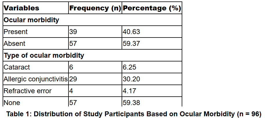
Non-neural granular cell tumor (NNGCT), a rare entity first described in 1991. The cell of origin is uncertain but histologically resembles conventional granular cell tumor, this is negative for S100 and lacks true nerve sheath differentiation. Here we present a case of 45-year-old female patient presenting with swelling over the right arm for 6 months which is diffuse. Cytosmear show large polygonal to spindled cells with pleomorphism, eosinophilic cytoplasm. Histopathological examination and multiple IHC markers helped us to rule out various differentials and diagnose as non-neural variant of Granular cell tumor.
Keywords: Granular, Nonneural, Soft tissue
Granular cell tumour (GrCTs) is a rare, benign, soft tissue tumour that likely arises from Schwann cells 1 . Primitive non-neural granular cell tumor(PNNGCT) is a variant of GrCT which is even more rare, low grade neoplasm of mesenchymal cells of unknown lineage 2, 3 . These tumors are most common in children and young adults, and often involves back and extremities 4, 5, 6 . Less than 100 cases of PNNGCT have been reported so far and these are usually benign. Immunohistochemically these tumors are S-100 protein negative 7 .
A 45-year-old female patient presented to surgical outpatient department with swelling over the right arm above elbow. Swelling was present since 6 months, sudden increase in swelling size since 1 month, associated with fever, No history of trauma. On examination, diffuse swelling noted over the right arm above elbow measuring 8x5cms, Firm to hard in consistency, mobile in horizontal plane, non-tender, skin over the swelling is tense.
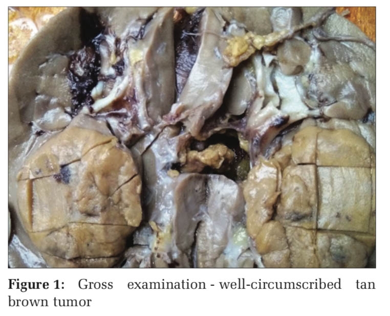
FNAC was done outside and reported as smears show moderate cellularity comprised of sheets, cohesive clusters, groups, singly scattered large polygonal to spindled cells having markedly pleomorphic hyperchromatic nuclei, abundant eosinophilic cytoplasm and features of pleomorphic sarcoma was considered and Further excision biopsy was advised. Surgery was done and specimen sent for histopathological examination. Received specimen well encapsulated globular grey white to grey brown soft tissue mass measuring 8x5.5x3cm. Cut section shows grey white areas with focal areas of necrosis (Figure 1).
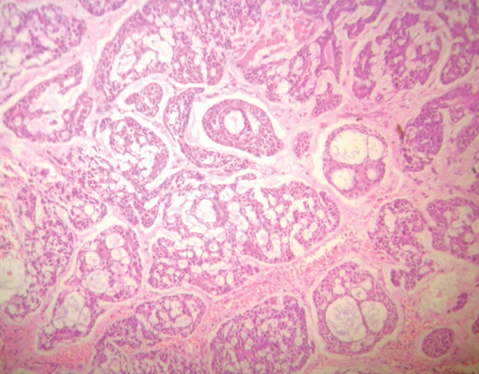
Section studied from the tumor tissue shows tumor arranged in solid sheets, in syncitial pattern and seperated by thin and thick fibrous septae. Individual cells are pleomorphic, polygonal with abundant granular eosinophilic cytoplasm with pleomorphic round to oval vesicular nuclei. Cytoplasmic membrane is indistinct at places. Many spindle shaped cells also noted. Large areas of necrosis seen. Atypical mitotic figures noted. Tumor is seen infiltrating adipose tissue (Figure 2). Sections from superior and lateral margins shows infiltration based on clinical and histological features we considered diagnosis as Malignant soft tissue tumor possibly Malignant granular cell tumor. To confirm the diagnosis, we have sent for Immunohistochemistry and we got s100, Desmin negative and positive for CD68 and NSE focally then we came to a conclusion that it’s a Granular tumor of non-neural origin which resembles Malignant Granular cell tumor histologically (Figure 3).


Granular cell tumors are a rare entity, representing only 0.5% of all soft tissue tumors. Of these, however, only a smaller percentage of 1-2% are malignant tumors, with a 40% mortality rate 7 . Fanburg-Smith et al. proposed six criteria for the histological diagnosis of malignancy; these include spindling, necrosis, increased mitotic activity, high nuclear cytoplasmic ratio, pleomorphism, and vesicular nuclei with large nucleoli. The presence of more than equal to three denotes as malignant tumor. In the present case, based on morphological features we thought differentials could be granular cell tumor, rhabdomyo tumor, Histiocytic tumor. To confirm IHC was done for s-100 it turned out to be negative, to rule out mesenchymal origin desmin was done which was also negative, then we performed IHC for CD 68 and NSE both of them were positive. When we went through the literature, we found a rare entity called S-100 negative Granular cell tumor which closely resemble conventional Granular Cell Tumor morphologically and histologically named as Primitive non neural Granular cell tumor. However, origin of this PNNGCT is unknown. Common site of presentation is oral cavity but, in our case, it is arm which is even rarer.
|
|
S-100 |
CD 68 |
NSE |
SMA |
DESMIN |
ALK |
CYCLIN D1 |
|
PNNGCT |
- |
+ |
+ |
- |
- |
+ |
+ |
|
GrCT |
+ |
+ |
+ |
- |
- |
- |
- |
|
Rhabdomyoma |
- |
- |
- |
+ |
+ |
- |
- |
|
Melanoma |
+ |
- |
- |
- |
- |
- |
- |
|
Epithelioid fibrous histiocytoma |
+/- |
+ |
- |
- |
- |
+ |
+ |
PNNGCT is rare tumor that was first described by Le Boit et al 8 . In 1991 on based on histological features and absence of reactivity towards S-100. Non neural granular cell tumors are well circumscribed lesion with vesicular nuclei, pleomorphism and increased mitotic activity. Cells are polygonal, oval or spindle with abundant eosinophilic cytoplasm. The diagnosis of certainty is made by histopathological and immunohistochemical examination 4 . These are usually negative for S-100, SOX10 which differentiate PNNGCT from conventional GrCT. They usually stain positive for CD63, CD68 and focal positive for ALK, NSE and CYCLIN D15 9 (Table 1). Although this is a rare tumor these are usually benign however malignancy cannot be ruled out and simple excision is alone sufficient in most of the cases.
This diagnosis of primitive non neural granular cell tumour should be considered in case of S-100 negative granular cell tumour to avoid incorrect interpretation and overt treatment. However, this unique tumor needs additional studies to understand and differentiate from other soft tissue tumors.
Subscribe now for latest articles and news.