Journal of Medical Sciences and Health
Year: 2022, Volume: 8, Issue: 2, Pages: 151-158
Original Article
Manisha Kumari1 , Sanjay Kumar Suman2 , Ruchi Gupta2 , Aishwerya Singh3 , Pragya Verma4 , Ashish Kumar Jha5
1Professor, Department of Radiodiagnosis, IGIMS, Patna, Bihar,
2Assistant Professor, Department of Radiodiagnosis, IGIMS, Patna, Bihar,
3Senior Resident, Department of Radiodiagnosis, Department of Gastroenterology, IGIMS, PMCH, Patna, Bihar,
4Assistant Professor, NMCH, Sasaram, Bihar,
5Additional Professor, Department of Gastroenterology, IGIMS, Patna, Bihar
Address for correspondence: Manisha Kumari, Professor, Department of Radiodiagnosis, IGIMS, Patna, Bihar.
E-mail: [email protected]
Received Date:25 January 2022, Accepted Date:10 July 2022, Published Date:05 September 2022
Background: Various imaging modalities like USG, CT, Invasive cholangiography has been used since long for the evaluation of the pathology of hepato-biliary-pancreatic duct. These techniques pose some limitations either due to bowel gas and obesity in USG, distal CBD calculus and isodense calculus (isodense to bile) in CT (also not used in patient with history of contrast allergy) and post-procedural complications (followed by invasive cholangiography like ERCP and PTC). MRCP is a non-invasive diagnostic technique for the direct visualization of the biliary ducts images similar to that produced in PTC (percutaneous trans-hepatic cholangiography) and ERCP. Also, the continuous expanding spectrum of therapeutic options, including radiological interventions (for palliative and curative) for the patients with biliary tree pathology requires the accurate assessment of the lesion visualized. Aims & objectives: Comparison of sensitivity, specificity and diagnostic accuracy between the MRCP/MRI and USG in patients with pancreatic and hepato-biliary pathology and it’s related complications. Materials & methods: The study was a retrospective, single institutional study done during the last two years (January 2018 – December 2019) at IGIMS, Patna, Bihar (A tertiary care hospital). Data was collected from the departmental record for USG findings, medical record for history and PACS (Picture archiving and communicating system) for MRCP findings. Hundred patients were included in our study. All the data were entered in the MS Excel sheet and were expressed as percentage and variables as required. Vassarstats software was used for the data analysis. Results: Benign lesions were common in the age range of 46-60 years (23%), followed by 31-45 years. Most of the malignant lesions were detected in the age range of 31-45 years of age group (13%). USG detected and MRCP detected lesions were benign in 63% and 62% of the cases and malignant in 34% and 35% of the cases respectively. There were overlapping findings present in most of the cases. Pancreatitis, cholecystitis and choledochal cysts were usually associated with biliary tree calculi. Overall sensitivity, specificity and diagnostic accuracy of MRI with MRCP sequence (97%, 85% and 92.75%) is greater than that of USG (92%, 80% and 85.5%). Conclusion: MRCP combined with other MRI sequences was superior to the USG in identifying benign and malignant pathology. However, ultrasound remains the primary investigating modality of choice. However, with use of recent advances of USG like harmonic imaging there is significant improvement in the lesion characterization. MRCP is the modality of choice of imaging investigation for the characterization of the lesion especially in obese patients or to choose the patients who are ideal candidate for the MRCP.
Keywords: Hepato-biliary-pancreatic pathology, MRCP
MRCP (magnetic resonance cholangio- pancreatography) with other MRI (magnetic resonance imaging) sequences like T1WI (T1 weighted images), T2WI (T2 weighted images) and DWI (diffusion weighted imaging) is a widespread, exciting and still emerging tool for the non-invasive assessment of the pancreatic and biliary ductal tree. 1 Obstructive jaundice is one of the most frequent forms of hepatobiliary disease and it faces several difficulties in diagnosis and management. Patients with jaundice are initially referred for the USG followed by other appropriate imaging modalities like CECT (contrast enhanced computed tomography), MRCP & ERCP (endoscopic retrograde cholangio-pancreatography). 2 Bowel gas, debris, fluid in duodenum and obesity make the assessment of CBD calculus or stricture difficult in the USG (ultrasonography).
MRCP is a non-invasive diagnostic technique for the direct visualization of the biliary ducts images similar to that produced in PTC (percutaneous transhepatic cholangiography) and ERCP. 3 MRCP is becoming the initial imaging tool for hepato-biliary pathology in some institutions and ERCP has been reserved for the therapeutic interventions like CBD calculus removal, CBD stent placement or ampullary dilatation. Common indications for the MRCP usually include unsuccessful ERCP, contraindication of ERCP and presence of biliary-enteric anastomosis. Advantages of MRCP are - non-invasive, no radiation, no requirement of anaesthesia, less operator dependent, better visualization of ducts proximal to obstruction, in combination of other MRI sequences (T1WI and T2WI) allows detection of extraductal disease, and it doesn’t have ERCP related complications like pancreatitis, infection, bleeding, bile duct injury/leak and even death sometimes. Disadvantages of MRCP are - decreased spatial resolution, imaging of non-distended biliary tree is usually difficult and intervention can’t be done at the same sitting.
USG has been still considered as the initial screening imaging modality in the study of biliary obstructive disease and other pathology, due to its accessibility, speed, ease of performance and low cost. 4 It is operator dependent and plays a key role in initial evaluation of the pathology. Traditional Computed Tomography (CT) scan is usually considered more accurate than USG to determine the specific cause and level of biliary obstruction. 5 But CT has some limitations and 10% CBD calculus which is radiolucent can be missed in CT scan.
Biliary strictures are also not visualized directly. It’s the diagnosis of exclusion, seen most often as abrupt termination of CBD without any mass lesion. Length and extent of the stricture is also difficult to determine by CT scan. Hence intravenous cholangiography (IVC), percutaneous transhepatic cholangiography (PTC) and endoscopic retrograde cholangio-pancreatography (ERCP) are used for the assessment of extent of stricture segment. 6
Due to limited role of the USG and CT scanning and invasive imaging technique of the IVC, PTC and ERCP; MRCP plays an excellent role in the assessment of the hepato-biliary tree and pancreatic duct for the diagnosis of the cause and level of obstruction. 7
Comparison of sensitivity, specificity and diagnostic accuracy between the MRCP/MRI and USG in patients with pancreatic and hepato-biliary pathology and it’s related complications.
The study was a retrospective, single institutional study done during the last two years (January 2018 – December 2019) at IGIMS, Patna, Bihar (A tertiary care hospital). The study protocol was approved by the Institutional Ethical Committee (IEC) for human research approval.
Data was collected from the departmental record for USG findings, medical record for history and PACS (Picture archiving and communicating system) for MRCP findings. Hundred patients were included in our study and their identity were kept anonymous.
Patient with all age and both sex and
Patient underwent both MRCP and USG examinations
Patients underwent only MRCP or only USG examinations
Follow-up case (to rule out duplication of data
MRI images with artefacts causing interpretations problem
3 plane LOC SSFSE — caliberation
Axial T2WI RTr PROPELLAR
T2WI FS RTr PROPELLAR
Coronal and Axial 3D MRCP RTr
Coronal T2WI SSFSE RTr
Axial DWI: Multi — b Value
2D Thick slab MRCP BH
Ax FS BH T1 FSPGR
COR FS FIESTA BH
Ax LAVA-Flex BH
Images were acquired in breath hold and the above parameters were individualized.
USG (Samsung H60 machine) performed routinely in this institute with maintaining standard protocol using a 3-5 MHz curvilinear probe in fasting state (overnight fasting).
All the data were entered in the MS Excel sheet and were expressed as percentage and variables as required. Vassarstats software was used for the data analysis.
Out of 100 patients, 55% of the study population was female and 45% was male.
|
Age (years) |
Number of patients |
Patients with Benign lesion (percentage) |
Patients with Malignant lesion / percentage |
|
<15 |
3 |
3 (3) |
0 (0) |
|
16-30 |
17 |
14 (14) |
3 (3) |
|
31-45 |
28 |
15 (15) |
13 (13) |
|
46-60 |
32 |
23 (23) |
9 (9) |
|
61-75 |
17 |
8 (8) |
9 (9) |
|
>75 |
3 |
1 (1) |
2 (2) |
|
Total |
100 |
64 |
36 |
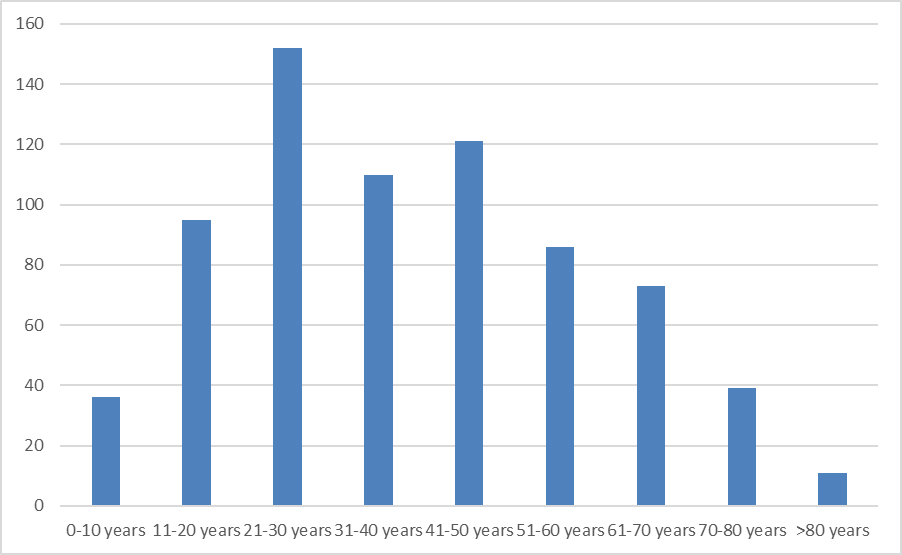
In this study, more patients were in the 46-60 years of age range constituting 32% followed by 31-45 years (28%) and 17% each in 16-30 years and 61-74 years of age range. Least number of patients were present at the extremes of age i.e. <15 years and >75 years of age (3% each).
Benign pathologies were found in 64% of patients and malignant pathologies were found in 36% of patients.
Benign lesions were most common in the age range of 46-60 years (23%), followed by 31-45 years and 16-30 years comprising 15% and 14% respectively.
Most of the malignant lesions were detected in the age range of 31-45 years of age group (13%), followed by 9% each in the age range of 46-60 years and 61-75 years.
|
Types of lesion |
USG detected lesion |
MRI/MRCP detected lesion |
|
Benign |
63 |
62 |
|
Malignant |
34 |
35 |
|
Indeterminate |
3 |
3 |
|
Total |
100 |
100 |
Benign lesions were more common than the malignant lesions. USG detected and MRCP detected lesions were benign in 63% and 62% of the cases and malignant in 34% and 35% of the cases respectively. Neither USG nor MRCP was able to differentiate the lesion in 3% of the cases. Among these 1 was benign organised sludge in distal CBD and 2 were of malignant (one was periampullary mass and another was pancreatic head mass) etiology that remained indeterminate in USG. Two lesions were benign (one was benign biliary stricture and another was choledocholithiasis) and 1 was malignant (mid CBD cholangiocarcinoma) that remained indeterminate in MRI/MRCP. These indeterminate cases were confirmed to be benign or malignant by ERCP or/and biopsies.
|
Diagnosis |
Number of patients |
US detection of lesion |
MRI/MRCP detection of lesion |
|
Benign |
64 |
|
|
|
Pancreatic divisum # |
5 |
0 |
5 |
|
Bifid configuration of MPD # |
1 |
0 |
1 |
|
Choledochal cyst # |
4 |
4 |
4 |
|
Calculus in CHD/CBD/ Cystic duct/GB/ intra-hepatic biliary tree/MPD # |
50 |
30 |
49 |
|
Pancreatitis+- sequale # - Acute - Chronic (+- calcific) |
8 |
6 |
8 |
|
3 5 |
3 3 |
3 5 |
|
|
Cholecystitis # |
4 |
3 |
4 |
|
Post-operative biliary injury/leak |
2 |
0 |
2 |
|
Benign biliary stricture # |
7 |
6 |
7 |
|
Intrahepatic lesion – cholangiolar/ pyogenic abscess, hydatid cyst |
11 |
7 |
11 |
|
10 1 |
5 2 |
10 1 |
|
|
Malignant |
36 |
|
|
|
GB mass # |
30 |
26 |
29 |
|
Cholangiocarcinoma – hilar and mid CBD # |
1 |
1 |
2 |
|
Periampullary and/or pancreatic head mass # |
5 |
5 |
6 |
|
Hepatic metastasis # |
3 |
3 |
2 |
# indicated the presence of more than one lesion/pathology in a patient
In this study, the most common type of lesion was benign, comprising 64%. There were overlapping findings present in most of the cases. Pancreatitis, cholecystitis and choledochal cysts were usually associated with biliary tree calculi. Pancreatic divisum, bifid configuration of the MPD and long cystic duct insertion were detected incidentally in patients with other disease processes like pancreato-biliary duct calculi, cholecystitis, GB mass, cholangiocarcinoma and in periampullary mass. MRCP/MRI accurately detected these in all patients while USG didn’t. Among malignant pathology, gall bladder mass was most common followed by periampullary and pancreatic head mass.
|
|
Overall MRCP detected lesion |
Overall USG detected lesion |
MRCP detected lesion (benign) |
MRCP detected lesion (malignant) |
USG detected lesion (benign) |
USG detected lesion (malignant) |
|
Sensitivity (%) |
97 |
92 |
94 |
100 |
94 |
90 |
|
Specificity (%) |
85 |
80 |
100 |
94 |
80 |
96 |
|
Diagnostic Accuracy (%) |
92.75 |
85.5 |
97 |
97 |
87.25 |
93 |
We found that overall sensitivity, specificity and diagnostic accuracy of MRI with MRCP sequence(97%, 85% and 92.75%) is greater than that of USG (92%, 80% and 85.5%). Sensitivity and diagnostic accuracy of detecting malignant lesions by MRI with MRCP were 100% and 97% respectively whereas the same for detecting benign lesions were 94% and 97%. Diagnostic accuracy of detecting benign lesions by MRI with MRCP (97%) was greater than that of USG (87.25%). Sensitivity of detecting benign lesions by USG was similar to that of MRI with MRCP (94% each).
In our study, 55% of the study population was female and 45% was male. Male to female (M/F) ratio was 0.82:1 which was comparable to those reported by Goyani et al 2 and Huis et al 8 being 0.76:1 and 0.69:1 respectively. However, in the study by Sharma et al M/F ratio was found to be 1.29:1. 9
More numbers of patients were present in 46-60 years (32%) followed by 31-45 years (28%). Least number of patients were present at extremes of age i.e. <15 years and >75 years (3% each). This result was comparable to the study conducted by Siva Prasad A et al 6 being biliary disease in 62% in the age group >40 year and 6% in the age group of less than 18 years and by Awadhesh Pratap Singh Kushwah in year 2015 who found the peak incidence in 51-60 year of age. 10 Nehal Diwanji et al in 2016 found that the peak incidence of pancreato-biliary disease was seen in 61-70 years of age. 11
Benign and malignant pathologies were found in 64% and 36% of patients respectively. Goyani et al 8 found that the benign and malignant lesions were present in 53.33% and 46.66% of the patients. Huis et al 8 observed that the benign and malignant lesions were present in 74.17% and 25.83% of patients. However, other studies by Siddique et al 12 , Sharma et al 9 , Moghimi et al 13 and Cheema et al 14 found that the malignant lesions were more common than the benign lesions.
Benign lesions were most common in the age range of 46-60 years (23%) and most of the malignant lesions were detected in the age range of 31-45 years of age group (13%). Goyani et al 2 and various other authors found that the most common age range in the benign and malignant lesions were 1-40 years and 41-70 years. This change in age pattern could be due to the changing trends of the disease with time.
USG and MRCP detected lesions were benign in 63% and 62% respectively and were malignant in 34% and 35% of cases respectively.
Neither USG nor MRCP was able to differentiate the lesion in 6 patients. These indeterminate cases were confirmed to be benign or malignant by ERCP and biopsies. Biopsy proven cases of benign and malignant lesions were 3 in number in each category. Biopsy proven cases of benign lesions were 3 in number that remained indeterminate in USG. One was benign (distal CBD organised sludge) and 2 were malignant (one was periampullary mass and another was pancreatic head mass) lesions among USG detected lesions in indeterminate category. Two were benign (one was benign biliary stricture and another was choledocholithiasis) and one was malignant (distal cholangiocarcinoma) among MRI/MRCP detected lesions in indeterminate category.
There was additional detection of hepatic cholangiolar abscess, postoperative biliary duct injury, small hepatic metastasis, distal CBD calculus or mass, accurate assessment of primary biliary confluence patency and various anatomic variants like pancreatic divisum, bifid configuration of the MPD and long cystic duct insertion by MRCP/MRI which USG failed to do. While choledochal cysts were detected both by USG and MRCP with 100% accuracy. USG showed its advantages in detection of the Mirizzi syndrome, gallbladder adenomyomatosis and chronic liver disease. MRCP combined with T1WI, T2WI and DWI sequences showed differentiation of distal cholangiocarcinoma and sludge which USG failed to do so.
Diagnostic accuracy and sensitivity of detecting malignant lesions and diagnostic accuracy of detecting benign lesions by MRI with MRCP was greater than that of USG 15 . Sensitivity of detecting benign lesions by USG was similar to that of MRI with MRCP. This could be explained by the use of graded compression technique and also use of THI (tissue harmonic imaging) which allows better visualization of the distal CBD; hence improved sensitivity and diagnostic accuracy by USG in our study. Ortega et al described that harmonic imaging, by improving contrast resolution, stressed the difference between the anechoic duct lumen and the surrounding soft tissues 16 .
Overall sensitivity, specificity and diagnostic accuracy of MRI with MRCP (97%, 85% and 92.75%) sequence were greater than that of USG (92%, 80% and 85.5%) in our study. Verma et al demonstrated the overall sensitivity, specificity and diagnostic accuracy were 80.77%, 95.83% and 88% for cases with benign condition on USG and the same for the malignant condition of biliary obstruction were 92.3%, 86% and 100% on MRCP 17 . Francesco SF et al found that the sensitivity, specificity and diagnostic accuracy of the USG were 16.67%, 97.29% and 78.62% and of MRCP were 90%, 94% and 93.13% respectively 18.


MRCP combined with other MRI sequences was superior to the USG in identifying benign and malignant pathology. MRCP is the modality of choice of imaging investigation for the characterization of the lesion specially in obese patients or to choose the patients who are ideal candidate for the MRCP. It also helps in the decision making regarding the management. It also allowed for the evaluation of the various anatomic variants of the pancreato-biliary system. However, ultrasound remains the primary investigating modality of choice. However, with use of recent advances of USG like harmonic imaging and 3-D/volume imaging there is significant improvement in the lesion characterization.
Subscribe now for latest articles and news.