Journal of Medical Sciences and Health
DOI: 10.46347/jmsh.v11.i1.24.435
Year: 2025, Volume: 11, Issue: 1, Pages: 111-113
Case Report
Bhavani Chikaraddi1 , A S Sanjana2 , M Dheemant3
1Post Graduate, Department of Dermatology, Venereology & Leprosy, BGS Global Institute of Medical Sciences, Bengaluru, Karnataka, India,
2Professor & HOD, Department of Dermatology, Venereology & Leprosy, BGS Global Institute of Medical Sciences, Bengaluru, Karnataka, India,
3Senior Resident, Department of Dermatology, Venereology & Leprosy, BGS Global Institute of Medical Sciences, Bengaluru, Karnataka, India
Address for correspondence:
M Dheemant, Senior Resident, Department of Dermatology, Venereology & Leprosy, BGS Global Institute of Medical Sciences, Bengaluru, Karnataka, India.
E-mail: [email protected]
Received Date:03 January 2025, Accepted Date:15 February 2025, Published Date:12 April 2025
Fibrous histiocytomas are mesenchymal tumors that can be benign or malignant. A cutaneous fibrous histiocytoma is a benign soft tissue tumour that can present as an indurated nodule anywhere in the human body. It is also known as dermatofibroma. A cutaneous fibrous histiocytoma usually presents as a small solitary firm papule or a nodule, seldom larger than 1 cm in diameter. We present here an atypical cutaneous fibrous histiocytoma presenting as a large keloid on the dorsum of hand in a 41-year-old female patient.
Keywords: Fibrous histiocytoma, Dermatofibroma, Benign mesenchymal tumour
Benign fibrous histiocytoma (BHF) or dermatofibroma (DF) is a mesenchymal tumor arising in cutaneous and non-cutaneous soft tissues. Cutaneous BHF usually originate in sun exposed areas of skin 1. They are mainly composed of fibroblasts and histiocytes forming a storiform growth pattern with overlying reactive epidermal changes 2. The pathogenesis of DF has been debated whether it is a neoplastic growth or a reactive process has not been settled. The fact that some DFs developed after insect bite and vaccination 3, 4 and the documentation of a history of trauma in 20% of DFs seem to favor a reactive process 5. However, DFs usually do not regress spontaneously with the exception of rare, documented instances 2. Therefore, the neoplastic theory cannot be dismissed entirely.
This article describes a case of atypical presentation of cutaneous fibrous histiocytoma masquerading as a large keloid on the dorsum of hand.
A 41-year-old female presented with a growing lump on the top of her right hand. She stated that the lesion presented without trauma or relevant history. The patient also noticed that the mass had grown in size since the past 1 year. The patient’s medical history was essentially unremarkable with the physical examination and radiographic analysis within normal limits. During inspection of the dorsum of the right hand, a red-brown, firm soft tissue mass measuring 2.5 × 3.0 × 0.5 cm was noted (Figure 1). The mass was freely movable, fluctuant, and presented with hyperkeratosis. The mass was nontender and presented with a flattened appearance. Following a local block, the lesion was excisionally biopsied and sent to the pathology department for confirmation.
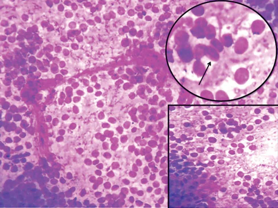
The results of the pathology report were consistent with cutaneous fibrous histiocytoma that showed skin lined by hyperplastic stratified squamous epithelium. Deeper dermis shows circumscribed tumour composed of benign spindle shaped cells arranged in storiform and pinwheel pattern. Figure 2).


A cutaneous fibrous histiocytoma is a common indurated nodule of benign origin composed of a mixture of fibroblastic and histiocytic cells 6. Alternatively, other terms such as dermatofibroma, fibrous xanthoma, atypical fibroxanthoma, sclerosing hemangioma, and nevoxanthoendothelioma have also been used 7. However, a simple classification that is currently used divides this lesion into two categories: The cutaneous fibrous histiocytoma refers to all superficial tumors of the skin regardless of appearance, whereas lesions penetrating into the subcutis, or deep structures are referred to as fibrous histiocytomas 8.
Most of these lesions arise spontaneously preceded by minor trauma. Numerous authors have documented mosquito bites as an inciting factor 9. Other injuries include scrapes, cuts, irritation from clothing, puncture wounds, and papulopustular lesions 5. The majority of these lesions occur between the ages of 20 and 40 years. Females are affected more often than males with no race cited as predominant 6.
Cutaneous fibrous histiocytomas commonly present as elevated and pedunculated lesions which may flatten over time. They can appear red, red-brown, and sometimes even black as a result of excessive deposits of hemosiderin. They usually range from a few millimeters to a few centimeters in diameter 6. Distribution is quite common on the lower extremities, especially in the anterior tibial area. These lesions, however, are rarely found on the foot, palms, and soles 8, 10.
A unique case of a histiocytoma is presented and was found to be inconsistent with the clinical features described in the literature; however, consistent with other aspects such as prevalence and onset. Although the lesion was chronic, there was no evidence of metastasis or malignancy. Excisional biopsy proved to be extremely effective. Currently, there have not been any recurrences or problems. Proper diagnosis and treatment plan with long term follow up helps in managing these tumors.
Subscribe now for latest articles and news.