Introduction
Marginal papular keratoderma is a heterogeneous group of palmoplantar keratodermas and refers to acrokeratoelastoidosis of costa (AKA) as well as focal acral hyperkeratosis of Dowd (FAH), which share similar clinical features of papules and plaques at the junction of palmar and dorsal skin of the hands or feet. They also show identical histologic epidermal alterations with subtle differences. They can be distinguished merely on the basis of absence of elastorrhexis in the latter. 1 Here, we report this unusual case of focal acral hyperkeratosis occurring at the age of 27-years -old female patient.
Case report
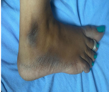
A 27-year-old female patient presented to dermatology OPD with brown to black coloured raised lesions on the borders of bilateral feet for the past 2 years. The patient denied any symptoms of itching, excessive sun-exposure, or trauma but the patient complained of hyperhidrosis of bilateral feet. Family history was insignificant, and lesions were not present anywhere else in the body.
On dermatological examination, there were multiple hyperpigmented, firm, discrete papules present on the lateral and medial borders of dorsum of bilateral feet (Figure 1, Figure 2). The differential diagnosis considered were marginal papular keratoderma, verruca plana, acrokeratosis verruciformis of Hopf, keratoelastoidosis marginalis and digital papular calcinosis.
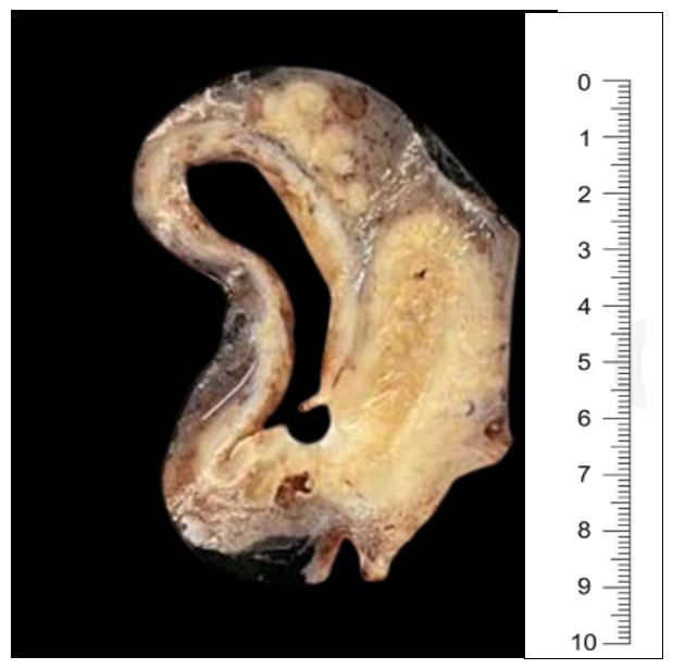
Further, biopsy of the lesion was done. Histopathological examination revealed, orthohyperkeratosis, mild hypergranulosis, acanthosis and the dermis showed no elastorrhexis giving a diagnosis of Focal acral hyperkeratosis which distinguishes from acrokeratoelastoidosis of Costa (Figure 3, Figure 4). The patient was treated with topical keratolytics like salicylic acid and retinoids, upon which patient noticed slight improvement.



Discussion
Palmoplantar keratoderma (PPK) is an umbrella term with a group of hereditary and acquired disorders characterised by thickening of skin over palms and soles. Under this major group of disorders, there is an entity called punctate palmoplantar keratoderma which are characterised by small keratotic papules in palms and soles, with infrequent involvement of their dorsal surfaces. Marginal papular keratodermas have emerged as a group within punctate palmoplantar keratoderma presenting with small firm keratotic papules along the borders of the palms and soles. Marginal papular keratodermas have further subtypes named acrokeratoelastoidosis (AKA) and focal acral hyperkeratosis (FAH), which have similar clinical presentation but differed by specific histologic changes of fragmentation of elastic fibres of the reticular dermis in AKA, but not in FAH. 2
FAH is a rare genodermatosis which was first reported in 1983 by Dowd et al. 3 It is hereditary and shows an autosomal dominant pattern of inheritance; however, it may be sporadic also. Our case doesn’t show any familial involvement proving it to be sporadic. It is more frequent among the African and middle eastern population with the onset of symptoms before 20 years of age in majority (80%) of the cases. 4 But here, we had encountered in an Indian population and the lesions had started after 25 years of age. There is no gender predominance in FAH. Clinically, it is characterised by small firm keratotic papules along the borders of hands and feet. They can be associated with itching, burning sensation on exposure to sun and sometimes hyperhidrosis. Occasionally, they are asymptomatic also. Here in our report, the patient had history of mild hyperhidrosis.
The pathogenesis of FAH remains largely unknown. Most of the PPKs have inherited genetic abnormalities encoding the structural components of keratinocytes, suggesting the analogous mechanism in FAH. 5 A report by Lee and Kim in 2007 demonstrated increased expression of proliferation markers (Ki-67 and PCNA) and differentiation marker (involucrin) in a patient with FAH. 6 This implies that the epidermal changes of FAH were mainly because of increased proliferation and differentiation of keratinocytes in the FAH lesion.
The histopathological findings of both FAH and AKA show common epidermal changes such as orthohyperkeratosis, mild hypergranulosis and acanthosis. The major distinguishing feature between the two is the lack of elastorrhexis in FAH.
The differential diagnosis for this clinical presentation included marginal papular keratoderma, verruca plana, acrokeratosis verruciformis of Hopf, keratoelastoidosis marginalis and digital papular calcinosis. 7 All these disorders exhibit similar clinical features; however, they differ in certain histopathological features. The different histological features are mentioned in Table 1.
|
Diagnosis |
Histological Findings |
|
Focal acral hyperkeratosis |
Focal hyperkeratosis, hyper- granulosis, acanthosis and with no elastorrhexis |
|
Acrokeratoelas-toidosis of Costa |
Focal hyperkeratosis, hyper- granulosis, acanthosis and with elastorrhexis |
|
Acrokeratosis verruciformis of Hopf |
Focal hyperkeratosis, acanth- osis, and papillomatosis (church spire configuration) |
|
Keratoelastoid-osis marginalis |
Hyperkeratosis, thick collagen, and elastic fibres |
|
Digital papular calcinosis |
Hyperkeratosis, acanthosis, and elastosis |
There is no effective treatment for this condition, yet many treatment modalities have been tried. 8 Topical keratolytics like salicylic acid, lactic acid, urea, retinoids, etc provide symptomatic relief to the patients. 9 Systemic retinoids like acitretin have also been tried in this condition. Our patient had slight improvement of lesions with topical keratolytics.
Conclusion
FAH has been described very rarely in the English literature and very few previous cases of FAH has been reported in Afro-Caribbean countries. To the best of our knowledge, only one case of FAH has been reported in Indian literature till now with the site of involvement being dorsum of bilateral hands. 10 Here, we are reporting this case of FAH over bilateral feet for its rarity among common dermatological practice and for its academic value. Although, the above condition is benign, it may cause psychological impact and cosmetic concern to the patient. However, further molecular studies may aid in improved understanding of FAH.

