Journal of Medical Sciences and Health
Year: 2022, Volume: 8, Issue: 2, Pages: 177-178
Case Report
K V Santosh1 , K N Manjunath2 , Vani Ravikumar1 , H N Ravikumar1
1Department of Pathology, RV Metropolis Diagnostic and Healthcare Pvt. Ltd, Bangalore
2Department of Plastic Surgery, MS Ramaiah Memorial Hospital, Bangalore
Address for correspondence:
K V Santosh, Department of Pathology, RV Metropolis Diagnostic and Healthcare Pvt. Ltd, Bangalore.
E-mail: [email protected]
Received Date:23 March 2022, Accepted Date:11 June 2022, Published Date:04 September 2022
Trichoadenoma is a rare benign tumor with differentiation to hair follicle. There are several reports of a melanocytic naevus seen in association with another benign adnexal tumor with hair follicular differentiation - trichoepithelioma. However, there is, to the best of our knowledge, only one case of a collision tumor of trichoadenoma with a melanocytic naevus - with an admixture of the two components. Trichoadenoma and trichoepithelioma are considered parts of a spectrum, with trichoadenoma being better differentiated than trichoepithelioma. Herein we present an unusual case of dual tumors - trichoadenoma and melanocytic naevi of the face. Unlike the previously reported case, the current case had simultaneous occurrence of trichoadenoma and naevi at different locations. The exact cause for the association of these tumors - trichoadenoma and melanocytic naevi - is uncertain.
Key Messages : Benign dermal adnexal tumors like trichoepithelioma and trichoadenoma may be found in association with melanocytic naevi.
Keywords: Dermal adnexal tumor, Trichoadenoma, Melanocytic Naevi
Trichoadenoma is a benign dermal adnexal tumor with differentiation towards hair follicle. While the association of melanocytic naevus with trichoepithelioma is well documented; there is, to the best of our knowledge, only one reported occurrence of a trichoadenoma with a melanocytic naevus - this case had an admixture of the two components within the same lesion. The current case has a combination of discrete lesions of trichoadenoma and melanocytic naevi involving the face. Unlike the previously reported case, the current case had no admixture of the components.
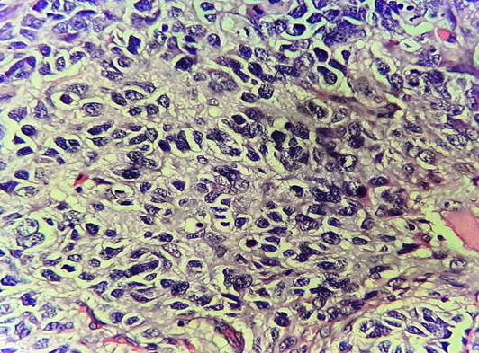
A 49-year-old male presented with history of multiple asymptomatic swellings over the face since 5 years. The lesions were located below the left eye, over the right nostril and in the region of the right side of chin. The patient did not recall the exact chronological sequence of occurrence of the 3 lesions, however, he had noticed that the swelling below the eye gradually progressed in size, while the others remained constant. He denied any other associated symptoms. On examination, all the swellings appeared pigmented. The lesion below the eye was located in infraorbital region, towards the root of the nose; measured 1x0.5 cm in size and had regular margins. The other 2 - one over the right ala of nostril and the other over the right chin region - measured 0.5 cm in diameter each. All three were excised. The microscopic section from the lesion near the left eye showed characteristic features of trichoadenoma (Figure 1), the dermis exhibiting several keratin horn cysts of varying caliber. These cysts contained luminal lamellated keratin and were lined by multilayered epithelium with eosinophilic cells rimmed by basaloid cells; with an intervening narrow granular layer. The other two lesions had the usual findings of dermal naevi comprising of dermal nests of nevoid melanocytes arranged in nests predominantly and demonstrating “maturation” pattern (Figure 2). Both the naevi lacked in junctional or intraepidermal component and were devoid of nuclear atypia or mitotic activity.


Benign dermal adnexal tumors with hair follicular differentiation include, among others, trichoblastoma, trichilemmoma, trichofolliculoma, trichoepithelioma, trichoadenoma and pilomatricoma. Trichoadenoma is a rarely encountered benign tumor with differentiation to hair follicle. The tumor presents as a nodule in the face most commonly, followed by the buttock, and measures 0.5 to 1.5 cm in diameter, with no gender preference. 1, 2 The clinical features may simulate a basal cell carcinoma or an epidermal cyst. 2 Despite different ontogenesis for dermal adnexal tumors and benign melanocytic naevi, there are several reports of a melanocytic naevus seen in association with trichoepithelioma. However, there is, to the best of our knowledge, only one case of a collision tumor of trichoadenoma with a dermal naevus has been reported; with an intimate admixture of the two components. 3 However, the current case had simultaneous occurrence of trichoadenoma and naevi at different sites. Trichoadenoma and trichoepithelioma are thought to represent two ends of a spectrum, 3 with cystic areas dominating the former and solid patterns predominant in the latter. 4 Trichoadenoma is considered to be better differentiated than trichoepithelioma, but not as well differentiated as trichofolliculoma. 5 Also, an IHC study using CK20 and Ber-EP4 suggested trichoadenoma to be a distinct tumor, albeit related to trichoepithelioma. 4 The occurrence of trichoepithelioma and naevi has been postulated as caused by epithelial induction of the adnexal tumor by the naevus. 6 It is uncertain whether the current example reflects a similar association.
Subscribe now for latest articles and news.