Journal of Medical Sciences and Health
DOI: 10.46347/jmsh.v12.i3.26.117
Year: 2026, Volume: 12, Issue: 3, Pages: 239-247
Original Article
Paridhi 1, Zeeba S Jairajpuri 2, Akanksha Malik 1, Mansi Agarwal 1, Sartaj Ali 1, Fouzia Siraj 1, Shruti Sharma 1
1Department of Pathology, ICMR- Centre for Cancer Pathology and National Institute of Child Health and Development Research, New Delhi, India.
2Department of Pathology, Hamdard Institute of Medical Sciences, New Delhi, India.
Address for correspondence: Shruti Sharma, Department of Pathology, ICMR- Centre for Cancer Pathology and National Institute of Child Health and Development Research, New Delhi, India.
E-mail: [email protected]
Received Date:19 March 2026, Accepted Date:13 June 2026, Published Date:19 August 2026

Histopathology relies on evaluating tissue sections to diagnose diseases, traditionally using xylene for deparaffinization. However, xylene poses health and environmental risks due to its toxicity. The study explores the efficacy of a 1.7% liquid dishwashing solution (DWS) as a safer, cost-effective and environmentally responsible xylene substitute in Haematoxylin & Eosin (H&E), histochemical and immunohistochemical staining. A retrospective, observational, comparative study was conducted over one year at a tertiary care centre with ethical approval. Hundred formalin-fixed paraffin-embedded (FFPE) tissue blocks from various organs were included. Two sections (4 µm thick) per block were stained using conventional xylene-based and xylene-free (XAF) methods. For histochemical stains (FF, PAS, MT) and IHC (15 antibodies), 20 blocks each were processed. The XAF protocol involved 1.7% DWS at 90°C for deparaffinization. Slides were assessed for nuclear and cytoplasmic staining, clarity, uniformity, and crispness. Statistical significance was determined using the Wilcoxon matched-pairs signed-rank test (P ≤ 0.05). Diagnostic staining quality was adequate in 95 (95%) of H&E slides with both methods. The XAF method showed superior nuclear staining, clarity, and crispness in 1 (1%), 3 (3%), and 2 (2%) cases respectively, while the conventional method had better uniformity in 8 (8%) cases. Histochemical and IHC results were comparable, with minimal variation. XAF reduced staining time significantly (30–35 minutes vs. 70–75 minutes) and lowered background staining in certain IHCs. A 1.7% DWS solution is a viable xylene alternative, offering improved safety, cost-efficiency, and faster processing without compromising diagnostic quality.
In histopathology ‘Diagnosis’ is referred to as the process of identifying and determining the nature and cause of a disease through complete evaluation of patient and review of the slides[1]. Tissues received in the histopathology laboratory undergo various stages of tissue processing to produce microscopic slides that are viewed by the pathologists for making a diagnosis of diseases. The bulk of daily diagnostic work in a pathology laboratory is of the paraffin section. Tissues embedded
in paraffin, which is similar in density to the tissue, can be sectioned at anywhere from 3 to 10 microns. The technique of getting fixed tissue into paraffin is called tissue processing, the main steps of which are dehydration and clearing[2].
The process of deparaffinization of the slides using Xylene is an important preliminary step before the staining process, which makes the tissue sections to take the stain properly. This makes Xylene an inevitable compound in histopathology laboratory due to its paraffin solvent action. Its high solvency factor allows maximum displacement of alcohol and renders the tissue transparent, enhancing paraffin infiltration; however, it is a toxic compound that is hazardous for human use and the environment in which it is disposed[1]. Therefore, a substitute that minimizes the use of Xylene in experiments, reduces tissue staining time and does not compromise its quality will be efficient for diagnostic
purposes and valuable for maintaining a safe laboratory environment.
Xylene was considered as the best clearing agent technically and also the safest alternative to dangerous chemicals like aniline oil, benzene, chloroform, toluene, dioxane etc. But there were great concerns about its safety. Exposure to xylene in a laboratory occurs during tissue processing, deparaffinization of tissue sections, cover slipping, cleaning tissue processors and recycling[2]. Toxic effects of xylene include acute neurotoxicity, cardiac and kidney injuries, hepatotoxicity, fatal blood dyscrasias, skin erythema, drying, scaling, secondary infection and also a carcinogenic effect.
Basic aim in any field of life sciences is to utilize eco-friendly chemicals which are nontoxic, less biohazardous, and are economical. After the hazardous effects of xylene became indisputable, many potential substitutes became available in order to make a xylene-free environment in laboratories, such as limonene reagents, aliphatic hydrocarbons, aromatic hydrocarbons, olive oil, vegetable oils, and mineral oil substitute. However, these chemicals were used to substitute xylene as a clearing agent during routine processing, while the exposure and handling of xylene is maximum during deparaffinization of the tissue sections. Liquid dish washing solution (DWS) which forms a part of the day-to-day household job has been experimented to dewax tissue sections by Falkholm et al in 2001[3]. It is a detergent liquid used to clean greasy utensils. These are anionic surfactants commonly used in detergent soaps and shampoos. Various laboratories have done pilot trials with successful outcomes; however, the results of these trials were varied with regards to individual parameters like nuclear staining, cytoplasmic staining and clarity. There were also other substitutes that showed promising results like diluted lemon water, mineral oil and hot distilled water[3].
Our study presents H&E, histochemical and Immunohistochemistry (IHC) staining technique that involved the use of easily available, nontoxic and eco-friendly diluted liquid dish washing solution (DWS) for deparaffinization and studied the efficacy of liquid detergent as an alternative to xylene and alcohol in Haematoxylin & Eosin and histochemical and IHC staining procedures and compare it with conventional method.
This was a retrospective, observational, and comparative study conducted over a period of one year at a tertiary care centre in New Delhi, India. The objective was to compare the diagnostic utility and staining quality of the conventional Haematoxylin and Eosin (H&E) staining method with the Xylene-Alcohol-Free (XAF) H&E staining method across a variety of tissues, histochemical stains, and immunohistochemical (IHC) markers.
Ethical clearance was obtained from the Institutional Ethics Committee prior to commencement of the study. All tissue samples were archival formalin-fixed paraffin-embedded (FFPE) blocks and used in accordance with institutional and national ethical standards for retrospective analysis.
A total of 100 FFPE tissue blocks were selected for H&E staining. These included a wide spectrum of tissue types: breast, lymph node, brain, bone, kidney, skin, thyroid, oropharynx, gastrointestinal tract, prostate, and urinary bladder. Inclusion criteria were well-preserved, adequately labelled FFPE blocks with sufficient tissue content. Blocks with damaged sections, inadequate tissue, or missing identifiers were excluded.
From each of the 100 blocks, two serial sections of 4 µm thickness were obtained, yielding 200 total sections. One section was stained using the conventional H&E method and the other with the XAF method for direct comparison.
Histochemical staining was performed on 20 FFPE blocks per stain for three commonly used special stains: Fite-Faraco (FF), Periodic Acid-Schiff (PAS), and Masson’s Trichrome (MT). Two 4 µm sections were prepared per block, totalling 120 sections (40 per stain).
For FF stain, 10 blocks each of leprosy tissue with bacillary indices 2+ and 6+ were selected.
For PAS stain, tissues with fungal infections, secreting adenocarcinomas, adenoid cystic carcinoma, and kidney-related conditions were chosen.
For MT stain, blocks included cases such as leiomyoma, collagenoma, sarcoidosis, regenerating nodules, angiomyolipoma, and urothelial carcinoma with muscularis invasion.
Immunohistochemistry (IHC) was performed using 15 commonly used antibodies in diagnostic pathology: Her2-neu, ER, PR, WT1, Pan-CK, Vimentin, CD34, p63, CK7, CK20, CD3, CD19, CD99, LCA, and Melan-A. For each antibody, 20 blocks were selected and two sections (4 µm each) were cut per block, resulting in 600 IHC-stained sections (300 by conventional, 300 by XAF method).
All staining procedures (H&E, histochemical, and IHC) were carried out simultaneously using both methods to reduce technical bias.
The XAF staining method involved deparaffinization using a 1.7% dishwashing solution (DWS) heated to 90°C. This was prepared by diluting 25 ml of commercially available Vim® liquid dishwashing soap (Hindustan Unilever Ltd.) in 1500 ml of distilled water. The detergent contained active ingredients including sodium laureth sulfate, sodium dodecylbenzene sulfonate, cocamidopropyl betaine, and non-ionic surfactants.
Following deparaffinization, standard H&E staining procedures were promptly applied. The process eliminated the need for xylene and alcohol, significantly reducing processing time to 30–35 minutes, compared to 70–75 minutes for the conventional method.
A) H&E Staining Evaluation
Each slide stained by either method was independently assessed by two blinded observers using the following five parameters:
Nuclear staining – Adequate (score 1), Inadequate (score 0)
Cytoplasmic staining – Adequate (score 1), Inadequate (score 0)
Clarity of staining – Present (score 1), Absent (score 0)
Uniformity of staining – Present (score 1), Absent (score 0)
Crispness of staining – Present (score 1), Absent (score 0)
The total score per slide (maximum of 5) was calculated. A total score of ≤2 was considered inadequate for diagnosis, while a score of 3–5 was considered adequate.
B) Histochemical Stains Evaluation
For FF, PAS, and MT stains, the evaluation included:
Specific localization of stain
Crispness of staining
Uniformity of staining
Each parameter was qualitatively assessed and compared between the Conventional and XAF methods.
C) IHC Stains Evaluation
For immunohistochemical stains, the slides were assessed based on:
Specific localization of antigen expression
Crispness of staining
Uniformity
Background staining
Evaluation was performed in a blinded manner by experienced pathologists.
Data were compiled in Microsoft Excel and analysed using GraphPad Prism software. The Wilcoxon signed-rank test was used for comparing paired non-parametric data. For each staining parameter, Z scores and P values were calculated. A p value < 0.05 was considered statistically significant.
|
|
Nuclear Staining | Cytoplasmic Staining | Clarity of Staining | Uniformity of Staining | Crispness of Staining | Scores Adequacy of Staining |
|---|---|---|---|---|---|---|
| Conventional Adequate | 98 | 98 | 95 | 98 | 95 | 95 |
| Conventional Inadequate | 2 | 2 | 5 | 2 | 5 | 5 |
| XAF Adequate |
99
|
96 | 98 | 90 | 97 | 95 |
| XAF Inadequate | 1 | 4 | 2 | 10 | 3 | 5 |
| Z value | -1.000 | -1.000 | -1.732 | -3.162 | -2.000 | 0 |
| P value | 0.317 | 0.317 | 0.083 | 0.002 | 0.046 | 1.0 |
| Significance | No significant difference between the two methods | No significant difference between the two methods | No significant difference between the two methods | Significant difference between the two methods | Significant difference between the two Methods | No significant difference |
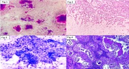
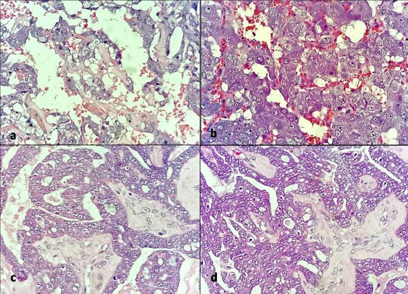
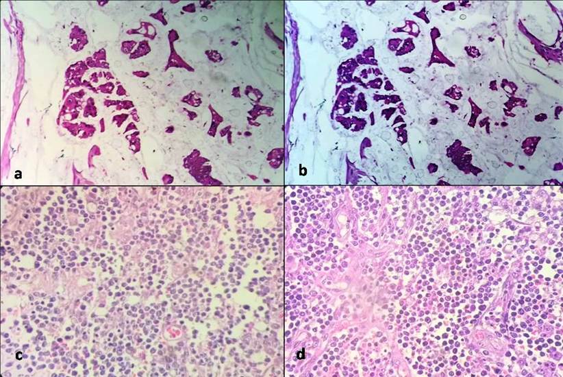
Slides stained by both the Conventional and Xylene Alcohol-Free (XAF) methods of H&E staining were found to be adequate for diagnosis in 95 (95%) cases. Nuclear staining was adequate in 98 (98%) cases with the Conventional method and 99 (99%) with the XAF method. While XAF showed slightly better nuclear staining in some cases, the difference was not statistically significant (P = 0.317, Z = -1.000). Similarly, cytoplasmic staining was adequate in 98 (98%) Conventional cases and 96 (96%) XAF cases, with no significant difference observed (P = 0.317, Z = -1.000) ([Fig. 1], [Fig. 2] and [Table. 1]).
Clarity of staining was found to be adequate in 95 (95%) of cases with Conventional staining and 98 (98%) with the XAF method. Although XAF showed better clarity in 3 (3%), the difference was not statistically significant (P = 0.083, Z = -1.732). In several cases, clarity appeared subjectively enhanced with XAF, particularly in the visualization of nucleoli and chromatin detail.
Uniformity of staining was adequate in 98 (98%) of cases using the Conventional method and 90 (90%) with XAF. In 8 (8%) cases, the Conventional method showed superior uniformity, and this difference was statistically significant (P = 0.002, Z = -3.162).
Crispness of staining was adequate in 95 (95%) Conventional cases and 97 (97%) XAF cases. Although the XAF method showed slightly better crispness in 2 (2%) cases, the difference was statistically significant (P = 0.046, Z = -2.000), favouring the Conventional method.
In terms of processing time, the XAF method required significantly less time (30–35 minutes) compared to the Conventional H&E method (70–75 minutes), making it more time-efficient for routine histological evaluation.
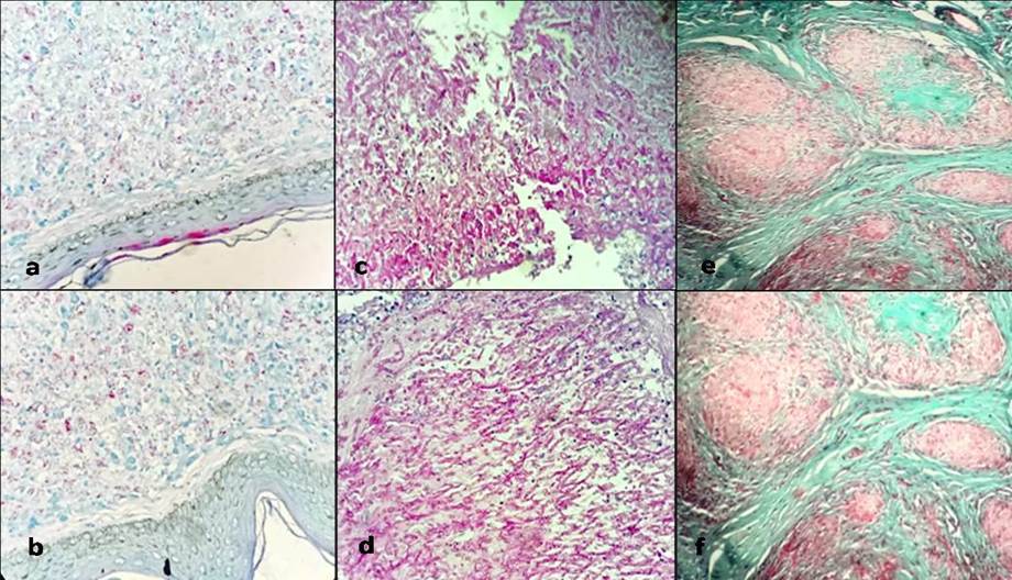
For histochemical stains, specific localization and uniformity of all three stains (PAS, FF and MT) both methods show similar results. Z-score (-1.01) and p-value (0.31) indicated no statistically significant difference between the conventional method (PAS Stain) and the XAF method. Similar results were observed for crispness of the staining of PAS and MT stain (p- value 0.31 and Z score (-1.01). The crispness of conventional method for FF was slightly lower than the XAF method (p value 0.14 and Z-score -1.45, again indicated no significant difference [Fig. 3].
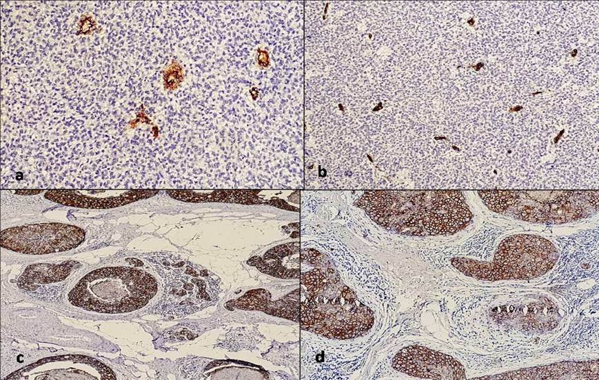
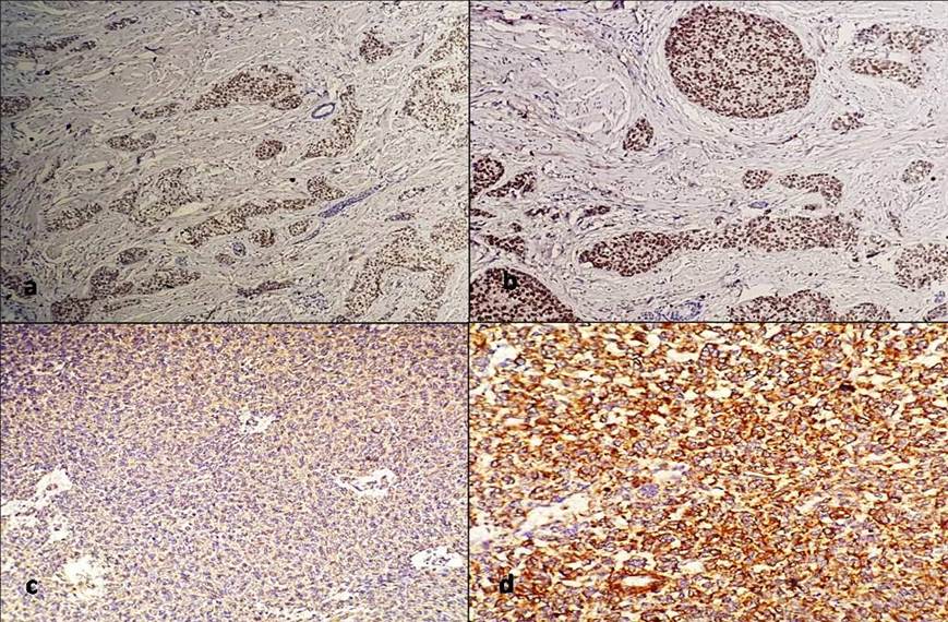
All the antibodies showed no difference between conventional and XAF method of staining, except for a few stains (ER, Vimentin, p63, CD19) where the conventional method shows a single absent case. However, p value was 1.0 for all the cases. Uniformity of staining was consistently higher in the XAF method compared to the conventional method (p-values ranged from 0.125 to 1), with most p-values being greater than 0.05, indicating no significant difference except for a few instances where p-values are close to 0.05 (e.g., Her2 neu, PR). The XAF method generally showed lower background staining compared to the conventional method. Several p-values were below 0.05 (e.g., PR, Vimentin), indicating a significant difference in background staining favoring the XAF method [Fig. 4]. Both methods showed high crispiness (p value 1.0), with the XAF method slightly outperforming the conventional method in almost all except vimentin, CD34, CK 20, Melan-A, where similar crispness were observed [Fig. 5].
Paraffin wax serves as a crucial embedding medium in routine H&E, Histochemical, and Immunohistochemical staining procedures. The staining process involves a series of essential steps, including dehydration, clearing, impregnation, and embedding. During these steps, paraffin sections are deparaffinized using xylene, followed by rehydration in graded alcohol. Xylene plays a pivotal role in these procedures and has become an integral component of histopathology laboratories. However, it is noteworthy that, currently, there is a lack of routine monitoring for the exposure of laboratory personnel to xylene. This widespread use without monitoring underscores the importance of evaluating and addressing potential occupational hazards associated with xylene exposure in histopathology laboratories[4, 5].
Xylene is widely used in histopathology lab for deparaffinizing the tissue section in H&E staining method, followed by rehydration in graded alcohol (methanol). The American Conference of Governmental Industrial Hygienists (ACGIH) has recommended the Biological Exposure Index (BEI) for various chemicals including xylene. However, in most of the developing countries, especially in India, we find no provisions for monitoring the exposure or any standardized methods for disposal of xylene[6, 7].
Xylene alternatives encompass even more toxic agents such as toluene, benzene, chloroform, cedar wood oil, methyl benzoate, and methyl salicylate. However, DWS stands out as a less toxic, cost-effective, and readily available alternative to traditional deparaffinizing agents like xylene for H&E staining procedures[3, 8].
The crucial aspect shared by all the substitutes was their requirement for high temperature (90°C) to be effective. Maintaining the temperature of diluted 1.7% liquid dishwashing liquid strictly at 90°C was imperative, slight deviation in temperature resulted in the washing away of tissue sections while temperatures below 90°C or if the sections were exposed for less time than recommended, the paraffin sections failed to deparaffinize completely, leaving residual wax in some areas. The significance of temperature in deparaffinizing paraffin sections was highlighted by Gyatri et al. They utilized 1.7% diluted dishwashing liquid at temperatures of 65/75/90°C for deparaffinization of sections instead of xylene, followed by H&E staining. The sections deparaffinized without xylene at 90°C were demonstrated to be as good as or even better than the conventional sections they were compared against[3, 9]. Liquid dishwashing detergents effectively deparaffinize wax through their surfactant action and pose fewer chances of toxicity to histotechnicians compared to the harmful effects of xylene[9].
The size and staining intensity of the nucleus are critical factors in disease evaluation. Proper staining of the nucleolus, nuclear membrane, and sharply defined condensed chromatin is of paramount importance[10]. There was no significant difference observed between the two methods.
| Ankle et al.[2] | Ramulu et al.[11] | Pandey et al.[8] | Ramaswamy et al.[5] | Current Study | ||
|---|---|---|---|---|---|---|
| Nuclear Staining | Conventional Adequate | 58 | 47 | 83 | 100 | 98 |
| Conventional Inadequate | 2 | 3 | 17 | 0 | 2 | |
| XAF Adequate | 59 | 48 | 90 | 100 | 99 | |
| XAF Inadequate | 1 | 2 | 10 | 0 | 1 | |
| Cytoplasmic Staining | Conventional Adequate | 56 | 46 | 76 | 100 | 98 |
| Conventional Inadequate | 4 | 4 | 14 | 0 | 2 | |
| XAF Adequate | 50 | 43 | 90 | 95 | 96 | |
| XAF Inadequate | 10 | 7 | 10 | 5 | 4 | |
| Clarity Of Staining | Conventional Adequate | 51 | 47 | 86 | 100 | 95 |
| Conventional Inadequate | 9 | 3 | 14 | 0 | 5 | |
| XAF Adequate | 53 | 48 | 90 | 75 | 98 | |
| XAF Inadequate | 7 | 2 | 10 | 25 | 2 | |
| Uniformity of Staining | Conventional Adequate | 42 | 46 | 88 | 98 | 98 |
| Conventional Inadequate | 18 | 4 | 12 | 2 | 2 | |
| XAF Adequate | 30 | 40 | 72 | 7 | 90 | |
| XAF Inadequate | 30 | 10 | 28 | 93 | 10 | |
| Crispness of staining | Conventional Adequate | 46 | 48 | 74 | 100 | 95 |
| Conventional Inadequate | 14 | 2 | 26 | 0 | 5 | |
| XAF Adequate | 50 | 44 | 95 | 23 | 97 | |
| XAF Inadequate | 10 | 6 | 5 | 77 | 3 | |
| Scores For Adequacy of Staining | Conventional Adequate | 53 | 47 | 84 | 100 | 95 |
| Conventional Inadequate | 7 | 3 | 16 | 0 | 5 | |
| XAF Adequate | 54 | 45 | 86 | 84 | 5 | |
| XAF Inadequate | 6 | 5 | 14 | 16 | 5 | |
| Total No of cases | 60 | 50 | 100 | 100 | 100 |
Furthermore, XAF staining demonstrated relatively superior adequacy in nuclear staining, particularly concerning the details of the nucleus and nucleolus, a finding consistent with the observations made by Ankle et al[2]. Our study employed Harris haematoxylin instead of Mayer’s haematoxylin for the XAF method. Remarkably, this substitution yielded equivalent results to those reported in other studies [Table. 2][11, 12].
In both XAF staining and conventional methods, 1% eosin Y was employed for cytoplasmic staining. Adequate similar cytoplasmic staining was observed in both the methods. Tap water with a pH of 7.02 was utilized both before and after the Eosin staining step (since Eosin is
acidic) to maintain alkalinity. Interestingly, no significant difference was observed between the two staining methods in terms of cytoplasmic staining. This finding aligns with a study conducted by Ramulu et al., where a few sections exhibited a bluish tinge in the cytoplasm [Table. 2][11].
Regarding the clarity of staining, it was found to be adequate in 95 (95%) cases with the two staining methods, however, not being statistically significant. This finding is consistent with the results reported in literature[2, 8, 11], and also indicates that XAF H&E staining is comparable to conventional H&E staining in terms of producing clear staining, our findings further support the notion that the XAF H&E staining method is equivalent to or even better than conventional H&E staining [Table. 2].
The uniformity of staining in the XAF sections was notably lower compared to the conventional H&E sections, where all sections showed uniform staining. Specifically, 90 (90%) XAF sections exhibited uniform staining. This slight difference indicates that conventional staining may yield better staining outcomes than XAF staining. Consistently, the issue of uniformity of staining with the XAF method was observed across all studies[2, 5, 8, 11]. After thorough investigation, potential causes of non-uniform staining such as chattering of sections, introduction of extraneous tissue, unclean blades, dirty microscopic lenses, thick sections, and moisture on cover slips were ruled out. Subsequently, it was determined that the xylene-free staining procedure is highly sensitive to temperature fluctuations. It was crucial to strictly maintain the diluted 1.7% liquid DWS-I and II, as well as the distilled water I and II, at precisely 90°C. Even a slight drop in temperature resulted in inadequate removal of wax from sections, while an increase in temperature led to the lifting and loss of sections from slides. Consequently, microscopic amounts of residual wax remained on the tissue sections, resulting in out-of-focus areas. Similar findings were observed in experimental studies conducted by Ankle et al. and Ramulu et al. [Table. 2][2, 11].
Regarding crispness, our observations were concurrent to that of Ankle et al. and Pandey et al., where XAF sections showed a significant difference in crispness as compared with conventional H &E. Ninety-six (96%) XAF sections revealed crisp staining as compared with 83 (83%) conventional H& E stain[2, 8]. The difference was significant, suggesting that XAF method is better over conventional method. However, Rumulu et al. found that conventional method was better than XAF method[11].
FF stain is employed to visualize acid-fast organisms like Mycobacterium leprae, which causes leprosy. In this staining method, leprosy bacilli appear red against a blue background. This distinctive coloration is due to the presence of mycolic acid; a waxy substance found in mycobacterial cell walls. The acid-fastness property of these organisms is directly related to the carbon chain length of the mycolic acid. Leprosy bacilli exhibit less resistance to both acid and alcohol. Consequently, alcohol is excluded from the hydrating and dehydrating steps of the staining process. Instead, 10% sulphuric acid is employed as a decolouriser in place of acid, ensuring optimal visualization of the acid-fast organisms while maintaining the integrity of the staining procedure[12, 13]. In the conventional method, sections are deparaffinized using a mixture of peanut oil and xylene. For our study, ten cases of leprosy with varying bacillary indices were selected. Staining was conducted using both conventional and XAF methods. Surprisingly, no discernible difference was observed in the positivity of bacilli between the two methods. Even with the XAF method, the bacilli displayed a bright magenta coloration and could be readily observed at 40x magnification. Notably, the magenta coloration was evident even in hair shafts, serving as an internal control. Remarkably, the identification of even single lepra bacilli was feasible, particularly in cases of borderline tuberculoid leprosy with a bacillary index of 2+. Furthermore, lepromatous leprosy cases with higher bacillary indices (6+) were vividly visualized. The staining exhibited uniformity with specific localization, presenting a very bright and crisp appearance. In fact, our findings indicated significantly improved results in terms of crispness compared to the conventional method.
PAS stain is used for detection of glycogen in tissues such as liver, cardiac and skeletal muscle on formalin-fixed, paraffin-embedded tissue sections. The glycogen, mucin, and fungi stained purple and the nuclei stained blue[14]. In our study, various tissues including skin tissue, kidney, and fungal infections such as aspergillosis, mycetoma, and histoplasmosis, as well as benign and malignant tumours such as secreting adenocarcinoma and adenoid cystic salivary gland carcinoma, were selected. Slides were stained using both conventional and XAF methods.
Notably, both conventional and XAF methods produced a bright magenta coloration of PAS-positive structures. Furthermore, specific localization, clarity, uniformity, and crispness were well appreciated in both methods. Based on these observations, we concluded that the XAF method serves as a viable alternative to the conventional method in PAS staining. This conclusion is supported by the fact that the XAF method allows for the appreciation of histological details while also being time-saving, cost-effective, and safe.
MT stain is used to differentiate between collagen and smooth muscle in tumours, and the increase of collagen in diseases such as cirrhosis[15]. In our study, various types of tissues including skin, kidney, as well as reactive benign lesions such as leiomyoma, collagenoma, chronic pyelonephritis, and sarcoidosis, along with malignant lesions like urothelial carcinoma infiltrating muscularis, were selected. The XAF slides exhibited brightness, crispness, and uniformity in staining comparable to conventionally stained slides. Notably, connective tissue, blood vessels, and collagen were well visualized in the XAF method, with specific localization of stain observed in various structures. Muscles and erythrocytes stained red, collagen stained blue, and nuclei stained black. Importantly, the muscle component in blood vessels appeared red in colour, and fibrotic areas were highlighted in green colour, particularly in cases of sarcoidosis where granulomas were separated by collagenous bands. The staining demonstrated uniformity with specific localization to respective tissues, appearing very bright and crisp.
A study by Henwood et al.[16] revealed that 51 of 55 antibodies showed equivalent staining with no significant differences in specificity, staining intensity, or background between conventional and hot detergent dewaxing methods. Additionally, improvements with hot detergent dewaxing for CD45RO and alpha fetoprotein were seen, while adverse effects were seen with CD10 and CD57. However, they found that conventional hydrocarbon dewaxing is significantly more expensive (839 times) compared to detergent dewaxing.16 Our study showed no significant differences in staining for most antibodies. Minor differences for ER, Vimentin, p63, and CD19 were seen where the conventional method showed a single absent case. Uniformity was even higher in the XAF method, though most p-values were >0.05, indicating no significant difference except for a few instances close to 0.05 (e.g., Her2 neu, PR). Background Staining was lower in the XAF method with significant differences for some stains (p-values <0.05), indicating improved clarity. Both methods showed high crispiness, with XAF slightly outperforming conventional methods in most cases, except for a few stains where both methods showed similar crispiness. Both the studies indicate that alternative methods to conventional dewaxing do not compromise staining quality and can be comparable or even better in certain aspects.
Faolain et al.[17] demonstrated that, aside from hexane, none of the other dewaxing agents were able to completely dissolve wax from tissue sections. Additionally, they observed that immunohistochemical results were significantly superior when sections were dewaxed with hexane compared to xylene[17].
Furthermore, DWS offers numerous advantages such as easy handling, cost-effectiveness (750 times cheaper than xylene), non-toxicity, and time-saving properties for staining. The use of 1.7% DWS in place of xylene is characterized by the absence of noxious odours in the laboratory and does not elicit irritation responses in laboratory workers. Therefore, DWS can be an appropriate alternative to xylene, maintaining the efficiency of staining without compromising quality.
The main limitations of the study were the XAF method’s sensitivity to temperature variations and the absence of long-term slide stability assessment, which may affect its broader applicability.
Dishwashing soap offers a viable and effective alternative to xylene in histopathological staining without compromising staining quality. When used in a diluted form, it facilitates efficient deparaffinization and supports staining procedures such as H&E, histochemical, and IHC techniques, producing results that are comparable to or even better than conventional methods. Slides processed with this method demonstrate clear and well-defined nuclear and cytoplasmic staining. In addition to its staining efficiency, this approach presents several practical benefits—it is non-toxic, non-flammable, cost-effective, easy to handle, and reduces both time and environmental hazards in the laboratory. Furthermore, Harris haematoxylin can be effectively used in place of Mayer’s haematoxylin in this method, yielding a similar quality of nuclear staining.
1. Biosafe substitute to xylene: A review. International Journal of Information Research and Review. 2016; 3 (6). Available from: https://www.ijirr.com/sites/default/files/issues-pdf/1220.pdf
2. A study to evaluate the efficacy of xylene-free hematoxylin and eosin staining procedure as compared to the conventional hematoxylin and eosin staining: An experimental study. Journal of Oral and Maxillofacial Pathology. 2011; 15 (2). Available from: https://doi.org/10.4103/0973-029x.84482
3. Xylene-Free Method for Histological Preparation: A Multicentre Evaluation. Laboratory Investigation. 2001; 81 (9). Available from: https://doi.org/10.1038/labinvest.3780335
4. Biosafe alternative to xylene: A comparative study. Journal of Oral and Maxillofacial Pathology. 2013; 17 (3). Available from: https://doi.org/10.4103/0973-029x.125199
5. A Study of Xylene Free Hematoxylin and Eosin Staining Procedure. Annals of Advance Medical Sciecnes. 2017; 1 (1). Available from: https://doi.org/10.21276/aams.1772
6. Bancroft JD, Gamble M. Theory and practice of histological techniques. 5th ed. London: Churchill Livingstone; 2002.
7. Significant changes in the amounts of neurotransmitter and related substances in rat brain induced by subacute exposure to low levels of toluene and xylene.. INDUSTRIAL HEALTH. 1983; 21 (3). Available from: https://doi.org/10.2486/indhealth.21.143
8. A comparative study to evaluate liquid dish washing soap as an alternative to xylene and alcohol in deparaffinization and hematoxylin and eosin staining. Journal of Laboratory Physicians. 2014; 6 (02). Available from: https://doi.org/10.4103/0974-2727.141504
9. Substitutes for xylene in histopathology and the role of temperature. Tropical Journal of Pathology and Microbiology. 2016; 2 (2). Available from: https://doi.org/10.17511/jopm.2016.i02.04
10. Sizing up the nucleus: nuclear shape, size and nuclear-envelope assembly. Journal of Cell Science. 2009; 122 (10). Available from: https://doi.org/10.1242/jcs.037333
11. Liquid dish washing soap: An excellent substitute for xylene and alcohol in hematoxylin and eosin staining procedure. Journal of Orofacial Sciences. 2012; 4 (1). Available from: https://doi.org/10.4103/0975-8844.99890
12. Efficacy of 1.5% Dish Washing Solution and 95% Lemon Water in Substituting Perilous Xylene as a Deparaffinizing Agent for Routine H and E Staining Procedure: A Short Study. Scientifica. 2014; 2014 Available from: https://doi.org/10.1155/2014/707310
13. Fite-Faraco staining in combination with multiplex polymerase chain reaction: A new approach to leprosy diagnosis. Indian Journal of Dermatology, Venereology, and Leprology. 2013; 79 (5). Available from: https://doi.org/10.4103/0378-6323.116740
14. Periodic acid-Schiff staining parallels the immunoreactivity seen by direct immunofluorescence in autoimmune skin diseases. North American Journal of Medical Sciences. 2016; 8 (3). Available from: https://doi.org/10.4103/1947-2714.179132
15. Masson Trichrome Stain Helps Differentiate Myofibroma from Smooth Muscle Lesions in the Head and Neck Region. Journal of the Formosan Medical Association. 2008; 107 (10). Available from: https://doi.org/10.1016/s0929-6646(08)60189-8
16. The application of heated detergent dewaxing and rehydration to immunohistochemistry. Biotechnic & Histochemistry. 2012; 87 (1). Available from: https://doi.org/10.3109/10520295.2010.518891
17. Raman Spectroscopic Evaluation of Efficacy of Current Paraffin Wax Section Dewaxing Agents. Journal of Histochemistry & Cytochemistry. 2005; 53 (1). Available from: https://doi.org/10.1177/002215540505300114
Subscribe now for latest articles and news.