Journal of Medical Sciences and Health
DOI: 10.46347/jmsh.v11.i2.24.353
Year: 2025, Volume: 11, Issue: 2, Pages: 197-202
Original Article
Neha Arya1 , Prashant Singh2 , Harendra Kumar3 , Chandrakanta3 , Jugendra Pal Singh Shakya4 , Pooja Nagayach2
1 Junior Resident, Department of Pathology, Sarojini Naidu Medical College, Agra, Uttar Pradesh, India,
2Associate Professor, Department of Pathology, Sarojini Naidu Medical College, Agra, Uttar Pradesh, India,
3Professor, Department of Pathology, Sarojini Naidu Medical College, Agra, Uttar Pradesh, India,
4Professor, Department of Surgery, Sarojini Naidu Medical College, Agra, Uttar Pradesh, India
Address for correspondence: Harendra Kumar, Professor, Department of Pathology, Sarojini Naidu Medical College, Agra, Uttar Pradesh, India.
E-mail: [email protected]
Received Date:29 January 2025, Accepted Date:01 March 2025, Published Date:07 July 2025
Background: Survivin is a cancer gene that is silenced in differentiated tissues, while overexpressed at high levels in vast majority of tumours. Some essential properties characterizing it as an ideal target involving apoptosis, promoting mitosis, stimulating vessel growth thus associated with poor prognosis, drug and chemo-resistance. The p53 gene is a tumour suppressor gene, defective p53 allows abnormal cells to proliferate resulting and cancer. The study aims to assess the expression of p53 and survivin in benign and malignant breast lesions and its prognostic importance. Materials and Methods: Total number of 102 patients were included in the study. Histopathologically confirmed 46 untreated female patients with Invasive breast carcinoma-NST and 56 female patients with fibroadenoma were used. Expression of survivin and p53 was evaluated using immunohistochemistry staining method. Results: In our study, we found 84.79% cases of invasive breast carcinoma-NST and 55.4% cases of fibroadenoma stained positive for p53, while 76.09% cases of invasive breast carcinoma and 71.42% cases of fibroadenoma stained positive for survivin. Conclusion: Increased expression of survivin and p53 noted in invasive breast carcinoma-NST as compared to fibroadenoma suggested that survivin and p53 are likely to contribute significantly to apoptosis resistance and may serve as therapeutic target that increase the effectiveness of conventional breast cancer therapy.
Keywords: Survivin, p53, Fibroadenoma, Invasive breast carcinoma-NST, Apoptosis, Immunohistochemistry
The breast cancer is most prevalent malignancy among women worldwide & manifests as palpable cancer in age groups of 40 and 50. 1 With an expected 2.3 million new cases (11.7%), female breast cancer has overtaken lung cancer as the most common cancer diagnosed, followed by lung (11.4%), colorectal (10.0%), prostate (7.3%), and stomach (5.6%) cancers. 2
Benign breast lesions are significantly more common than malignant ones. 3 Frequency of malignant diseases rises after menopause. The rise in benign breast lesions occurs during the second decade of life. 4 A fibroadenoma is usually unilateral & painless benign solid breast tumour, frequently affects women between the age of 14 and 35 years. Postmenopausal women are less likely to have fibroadenoma because they shrink after menopause. 5
Invasive breast carcinoma (IBC) comprises approximately 80% of breast cancers and most common variant of it is Invasive breast carcinoma of no special type (IBC-NST). Second most common type is lobular carcinoma, representing 15% of all breast cancer 6 followed by medullary, mucinous, comedo-carcinoma, Paget’s disease, papillary, tubular, and inflammatory carcinoma.
It is necessary to differentiate benign lesions from in situ and invasive breast cancer to establish the most appropriate therapy. 7 Mostly breast lesions are classified on the basis of morphological criteria but cases with equivocal morphology require ancillary studies e.g. immunohistochemistry; to reach at precise diagnosis, to know the prognostic behaviour & response to the therapy. 8, 9
Survivin is an anti-apoptotic protein that belongs to the inhibitor of apoptosis protein, having a unique structure and function. 10 Survivin overexpression in cancer may destroy this apoptotic checkpoint and permit abnormal mitotic proliferation of altered cells. 11
p53 gene is located on chromosome 17 [17p13.1], also known as TP53- Tumour Protein 53, which regulates the cell cycle and functions as a tumour suppressor protein. A p53 mutation leads to overexpression of survivin, which prevents apoptosis and results in tumours. 12, 13
In present study we have evaluated the expression of p53 & Survivin in both benign and malignant breast lesions.
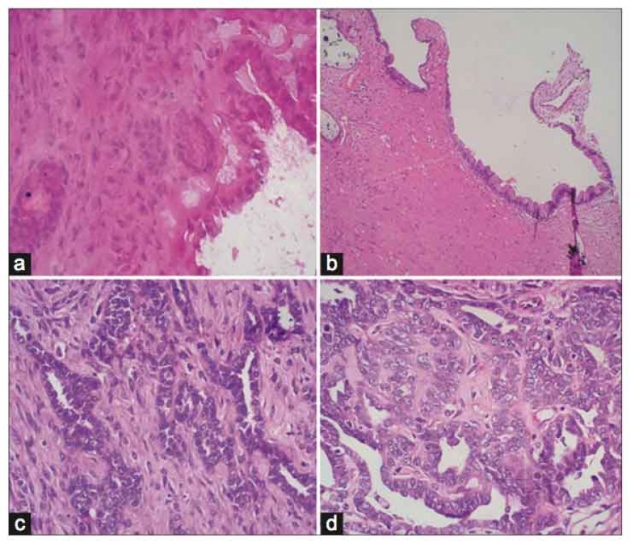
The cross-sectional study was conducted in the Department of Pathology of our Institute. The study was conducted over period of 18 months from September 2022 to May 2024. Ethical approval was obtained from the institutional ethical committee (ref: SNMC/IEC/2024/261). A total of 102 cases (56 cases of benign lesion and 46 cases of malignant lesions) of surgically resected mastectomy, lumpectomy and biopsy of breast specimens were included in the present study. After receiving the samples, gross inspection was done, and the size and appearance of the tumour were documented. After incising (bread loafing), the specimens were fixed in 10% formal saline for 12-24 hours. Tissue sections from representative areas were then submitted for further processing and paraffin wax blocks were made. Section of 3-4 µm were made from the paraffin wax blocks and stained by routine H&E dye, then examined under a light microscope. A histological diagnosis was made using modified Bloom-Richardson histological grading. Another section of 3-4µm was used for IHC for p53 and Survivin. the representative sections from all the cases were studied. The sections were evaluated at high power (400X microscopic field; objective 40X, eyepiece 10X), considering a high -power microscopic field harboured 100 stromal cells.
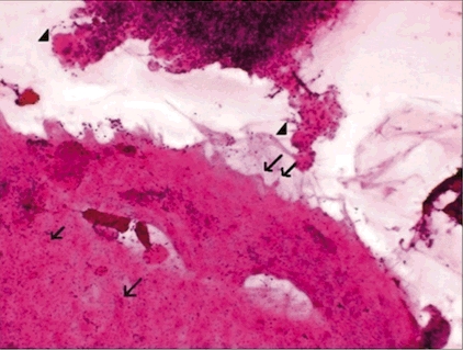
Positive control sections of p53 and Survivin were obtained from sections of brain tumour (diagnosed cases of astrocytoma- p53 mutant) and colon mucosa respectively. Positive histological reaction for Survivin antibodies used was visualized as cytoplasmic and in few cases nuclear brown staining, whereas for p53 positivity appeared as nuclear brown stain. A semi quantitative scoring system was used to score immunohistochemical positivity. The intensity of immunohistochemical staining for statical analysis was graded on a scale of 0 to 4.
The level of expression was scored as follows:
0 = negative, less than 5% of cells staining
1+ = weak staining, between 6% and 25% of cells staining
2+ = moderate staining, between 26% and 50% of cells staining
3+ = medium strong staining, between 51% and 75% of cells staining
4+ = strong staining, more than 75% of cells stains.
Inclusion criteria: All breast tru-cut biopsy, incisional biopsy, lumpectomy, and mastectomy specimens of patients with neoplastic (benign & malignant) aetiology confirmed on histopathology were included in the study.
Exclusion criteria: Inadequate and autolysed tissue and patient with metastatic tumours from systemic malignancies, all inflammatory/infective lesions and cases other than fibroadenoma and Invasive Breast Carcinoma -NST were excluded from the study.
The study included 102 cases in total, of which 46 had invasive breast carcinoma-NST and 56 had fibroadenoma. Most cases (26 cases) of fibroadenoma were noted in 2nd & 3rd decade. Most of the IBC cases (28 cases) were diagnosed in the 41-50 years age group, out of which 31.0% were IBC Grade-1, 37.9% having IBC Grade-2, 10.3% having IBC Grade-3. The statistical difference between the different age groups in terms of frequency distribution for invasive breast carcinoma (IBC-NST)-Grade 1, IBC-Grade 2, IBC-Grade 3, IBC-Grade NOS was statistically significant (Figure 1).

In our study, 54.9% of the lesions were diagnosed as fibroadenoma, 8.8% were IBC-Grade 1 lesions, 17.6% were IBC-Grade 2 lesions, 6.9% were IBC-Grade 3 lesions and 11.8% were IBC-NOS. Mean age of fibroadenoma and IBC grade 1, 2, 3 & NOS was found statistically significant, which was 0.001 (Table 1).
|
|
Mean |
Std Dev |
Std Error |
P value |
|
Fibro adenoma (54.9%) |
23.053 |
6.910 |
0.923 |
0.001 (Sig) |
|
IBC-Grade 1 (8.8%) |
47.111 |
3.100 |
1.033 |
|
|
IBC-Grade 2 (17.6%) |
52.388 |
10.273 |
2.421 |
|
|
IBC-Grade 3 (6.9%) |
49.285 |
5.794 |
2.189 |
|
|
IBC-NOS (11.8%) |
49.916 |
8.836 |
2.550 |
Out of 46 cases of Invasive breast Carcinoma-NST, 21.74% were having more than 75% tubular component, 67.39% were having 10-75% tubular component and 10.87% were having less than 10% tubular component. Out of 46 cases of Invasive breast Carcinoma-NST, 32.61% cases showed small, uniform cells, 56.52% showed moderate pleomorphism and 10.87% showed marked nuclear pleomorphism. Among the 46 cases of IBC-NST, 6.52% had 0-5/10 HPF mitotic count, 76.09% showed 6-10/10 HPF and 17.39% showed more than 10/10HPF mitotic count.
Lymph nodes were identified in the total of 27 cases out of total 46 cases of IBC-NST. Out of these 27 cases 10 (37.03%) were negative for metastasis and 17 (62.97%) were positive for metastasis.
Among the patients with fibroadenoma, 55.4% of the patients showed positive p53 Expression, whereas among the patients with Invasive breast Carcinoma-NST 84.79% of the patients showed positive p53 expression. Grade -1 lesion patients were having higher percentage of negative expression, whereas lesions with Grade-2, 3 and NOS, there was higher percentage of the patients having positive expression. The correlation of expression of p53 with different type of lesions was statistically significant with p value of 0.012. Among the patients with fibroadenoma, 71.42% of the patients showed positive Survivin expression whereas among the patients with Invasive breast Carcinoma-NST, 76.09% of the patients showed positive Survivin expression. Grade -1 lesion have higher percentage of negative expression, whereas among the patients with Grade-2, 3 and NOS higher percentage of the patients were having Positive expression. The correlation of expression of Survivin staining with different type of lesions was statistically significant with p value of 0.001 (Table 2).
|
|
p53 |
Survivin |
||
|
Negative |
Positive |
Negative |
Positive |
|
|
Fibroadenoma (total cases = 56) |
25 |
31 |
16 |
40 |
|
44.6% |
55.4% |
28.58% |
71.42% |
|
|
|
|
|
|
|
|
IBC-NST (total cases = 46) |
7 (13.21%) |
39 (84.79%) |
11 (23.91%) |
35 (76.09%) |
|
IBC Grade -1 (9 cases) |
3 |
6 |
6 |
3 |
|
33.3% |
66.7% |
66.67% |
33.33% |
|
|
IBC Grade -2 (18 cases) |
2 |
16 |
2 |
16 |
|
11.1% |
88.9% |
11.12% |
88.88% |
|
|
IBC Grade -3 (7 cases) |
0 |
7 |
0 |
7 |
|
0% |
100.0% |
0% |
100% |
|
|
IBC-NOS (12 cases) |
2 |
10 |
3 |
9 |
|
16.7% |
83.3% |
25% |
75% |
|


According to latest WHO breast 5th edition, terminology of IDC has been changed to Invasive Breast Carcinoma and the most common variant of it is Invasive breast carcinoma of no special type (IBC-NST). 14, 15
Survivin is a multifaced protein, which is nearly non-existent in most terminally differentiated adult tissues and frequently expressed in many different types of cancer in humans. It is also known that this antiapoptotic protein is located in the nucleus and cytoplasm. 10 Survivin binds to the microtubules of the mitotic spindle and is expressed in a controlled manner during the G2/M phase of the cell cycle. The antiapoptotic properties of survivin are lost when survivin-microtubule connections are disrupted, and caspase-3 activity is elevated during mitosis. This apoptotic checkpoint may be destroyed by survivin overexpression in cancer, allowing altered cells to proliferate abnormally through mitosis. These findings suggest that survivin may be able to block the G2/M phase default induction of apoptosis. In addition, it was shown that cultured breast cancer cells expressed increased survivin when exposed to chemotherapy, possibly as a defence against apoptosis. Immunohistochemical staining reveals that anti-survivin mAb 8E2 specifically reacts with breast carcinoma cells, with positive staining of the cytoplasm of cancer cells whereas, no expression of survivin seen in adjacent normal tissues. 11
Defective p53 allows abnormal cells to proliferative and resulting in cancer. Tumour suppression is severely reduced, if p53 damaged. 12, 16 In normal forms, it promotes apoptosis and controls the cell cycle as a result. In its mutant state, it promotes the growth of tumours by blocking apoptosis and losing control over the cell cycle. One of the most prevalent (approximately 50%) abnormalities in primary human cancer is overexpression of the nuclear phosphoprotein p53, which is thought to be caused by a single mutation in a highly conserved area of the p53 gene that results in the encoding of a mutant, more stable protein. 13, 17
In our study 56 (54.9%) cases belonged to fibroadenoma and 46 (45.9%) cases belonged to Invasive breast carcinoma-NST which were different from the study population of Nath D et al 18 , F Kayaselcuk et al 19 , Raj S et al 16 , Adamkov M et al 20 , Ranade K J et al 17 , Adamkov M et al. 10 This may be due difference in population selected in different areas.
|
Studies |
Total number of cases |
Most common age group |
Maximum tubular component |
Maximum nuclear grade |
Maximum mitotic count |
P53 expression |
Survivin expression |
||||
|
|
FA
|
IBC
|
FA
|
IBC
|
In IBC cases (%)
|
In IBC cases
|
N.A.
|
FA Cases (%)
|
IBC Cases (%)
|
FA Cases (%)
|
IBC Cases (%)
|
|
Nath D et al. 18 |
41 |
34 |
11-20 yr |
41-50 yr |
10-75% |
Moderate pleomorphism |
6-10/10hpf |
20 (50%) |
29 (85.30%) |
20 (37.50%) |
27 (79.40%) |
|
Raj S et al. 16 |
41 |
34 |
11-22 yr |
41-50 yr |
10-75% |
Moderate pleomorphism |
Not reported |
5 (12.9%) |
29 (85.30%) |
15 (37.50%) |
27 (79.40%) |
|
Our study |
56 |
46 |
11-20 yr |
41-50 yr |
10-75% |
Moderate pleomorphism |
6-10/10hpf |
31 (55.4%) |
39 (84.79%) |
40 (71.42%) |
35 (76.09%) |
First limitation of the study is smaller sample size and second is in-availability of all morphological variants of breast lesions, so more research, with all morphological variants of breast lesions, bigger sample size and long term follow up, are required to validate the results.
We concluded that apoptosis resistance in Invasive breast carcinoma patients is probably significantly influenced by the increased expression of p53 and survivin in these patients in contrast to fibroadenoma and may serve as therapeutic target that could increase the effectiveness of conventional breast cancer therapy. To find out whether p53 and survivin may be employed as prognostic markers, however, large sample size studies with clinical follow-up data are needed.
Funding: Nil
Conflict of Interest: Nil
Subscribe now for latest articles and news.