Journal of Medical Sciences and Health
Year: 2023, Volume: 9, Issue: 2, Pages: 229-232
Case Series
Sneha Kakoty1 , Dhaval Jetly2 , Paheli Maru1 , Anurag Saha1 , Varnika Rai1
1DM resident, Department of Oncopathology, GCRI, Ahmedabad, Gujarat, India,
2Associate Professor, Department of Oncopathology, GCRI, Ahmedabad, Gujarat, India
Address for correspondence:
Dhaval Jetly, Associate Professor, Department of Oncopathology, GCRI, Ahmedabad, Gujarat, India.
E-mail: [email protected]
Received Date:29 January 2023, Accepted Date:07 August 2023, Published Date:26 September 2023
Foamy gland carcinoma (FGC) is a distinct histological variant of prostatic acinar adenocarcinomas. It was first described by Epstein and Nelson in 1996. It is characterized by abundant xanthomatous cytoplasm, pyknotic nuclei, and intermediate Gleason grade. However, FGC with a high Gleason grade exists. FGC is found admixed with conventional acinar adenocarcinoma in 17 to 22% of needle biopsies and 13 to 22% of radical prostatectomy specimens and only rarely found in pure form. We report six cases of pure foamy gland carcinoma of prostate. The mean age in our study was 62.83 years and 5 out of the 6 cases presented with an elevated serum PSA level. Histopathological examination of our cases showed tumor cells with abundant foamy cytoplasm and low nucleo-cytoplasmic ratio. This deceptively benign-looking morphology can mimic nonneoplastic prostatic glands, Cowper’s gland, mucinous metaplasia, and xanthomatous prostatitis and hence often pose diagnostic challenges, especially in core needle biopsies. PNI was detected in two of our cases. PSA and AMACR staining was positive in all our cases (100%). p63 Staining was negative in all four cases where it was performed. Three, two, and one of our cases had a Gleason score of 7, 8, and 6 respectively. The prognosis depends on the Gleason score and the presence or absence of PNI or extraprostatic extension. Three of the cases presented with bony metastasis. We report this case series in view of the rarity and also to raise awareness of this entity which is often missed on small biopsies.
Keywords: Foamy gland carcinoma, Gleason score, Prostate
Foamy gland carcinoma (FGC) is a distinct histological variant of prostatic acinar adenocarcinomas. It was first described by Epstein and Nelson in 1996. It is characterized by abundant xanthomatous cytoplasm, pyknotic nuclei, and intermediate Gleason grade. 1 However, FGC with a high Gleason grade exists. Due to its deceiving benign histologic appearance, it often poses diagnostic challenges, especially in core needle biopsies. FGC is found admixed with conventional acinar adenocarcinoma in 17 to 22% of needle biopsies and 13 to 22% of radical prostatectomy specimens and is only rarely found in pure form. 2, 3 We report six cases of pure FGC of prostate because of its rarity and to raise awareness of this entity which is often missed on small biopsies.
A 58-year-old man presented with difficulty in urination. Ultrasonography (USG) showed a polypoidal lesion involving the base of bladder. Serum prostate-specific antigen (PSA) levels were 12.2 ng/ml. TURP was performed. Microscopically, it showed tumor cells with foamy cytoplasm arranged in infiltrative irregular and discrete glandular patterns. Perineural invasion was absent. Immunohistochemistry (IHC) showed positivity for PSA and AMACR and negativity for CK 7, CK20, beta-catenin, and CDX2 and was diagnosed as FGC with Gleason score 7 (4+3) and grade group 3.
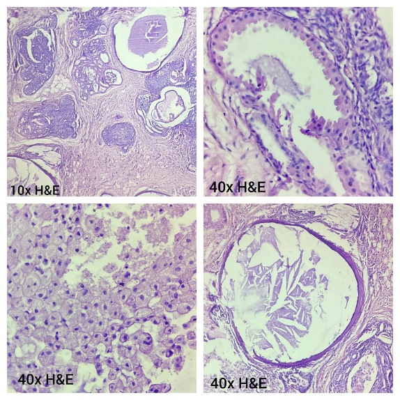
A 61-year-old man, a known case of prostatic carcinoma with a fracture right femur underwent Figure 1 a and b). Perineural invasion was noted (Figure 1 c). IHC showed positivity for AMACR, PSA, and GATA3 and negativity for CK7, CK20, and p63 (Figure 2 a to c). Diagnosis of FGC with a Gleason score of 8 (4+4) and grade group 4 was made.


A 74-year-old man underwent TURP following an MRI which showed an enlarged prostate gland with hyper-enhancing focus in right peripheral zone. Sclerotic metastatic lesions were noted in multiple vertebral bones. Serum PSA was 73.91ng/ml. Microscopic features were the same as in case 1. Perineural invasion was absent. IHC showed positivity for PSA and AMACR and negativity p63. It was diagnosed as FGC with a Gleason score of 7 (4+3) and grade group 3. A bilateral orchidectomy was performed.
A 50-year-old man presented with backache and difficulty in urination. MRI showed multiple vertebral bony metastases. USG showed enlarged prostate. Serum PSA was 1292ng/ml. Trucut biopsies from both sides of prostate were performed. Microscopic features were the same as in case 2. Perineural invasion was absent. IHC showed positivity for AMACR and PSA, and negativity for p63. It was diagnosed as FGC with a Gleason score of 8 (4+4) and grade group 4. He received palliative radiotherapy.
A 64-year-old man came for a review of his previous biopsy done for enlarged prostate detected on MRI. Serum PSA and skeletal survey were normal at the time of review. Microscopic features showed tumor cells with foamy cytoplasm arranged in discrete glandular patterns. Perineural invasion was absent. IHC showed positivity for AMACR and PSA, and negativity for p63. It was diagnosed as FGC with a Gleason score of 6 (3+3) and grade group 1.
A 70-year-old man underwent a radical prostatectomy and came for review. His serum PSA at the initial presentation was 95 ng/ml. MRI showed an infiltrative lesion on right side of prostate. No skeletal metastasis was detected on scintigraphy. Microscopy showed morphology similar to case 1. Perineural invasion and lymphovascular invasion were present. It was diagnosed as FGC with a Gleason score of 7 (3+4) and grade group 2. Both the seminal vesicles and six out of ten pelvic lymph nodes were involved by the tumor (Figure 3 a to c) Pathological stage was found to be pT3bN1.

Foamy gland carcinoma (FGC) is a distinct histologic variant of prostatic acinar adenocarcinoma. Epstein and Nelson first described this variant in 1996 which is characterized by abundant xanthomatous cytoplasm and pyknotic nuclei. 1 The foamy nature of the cytoplasm is due to the presence of numerous intracytoplasmic vacuoles which is devoid of lipid or neutral mucin. 4
The mean age for FGC is the same as usual for acinar adenocarcinoma which is 65 years (range 50-78 years). The mean age in our study was 62.83 years. They present with an elevated serum PSA level which was also observed in our study.
Histopathological examination of all our cases showed tumor cells with abundant foamy cytoplasm and low nucleo-cytoplasmic ratio. This deceptively benign-looking morphology can mimic nonneoplastic prostatic glands, Cowper’s gland, mucinous metaplasia, and xanthomatous prostatitis. Neoplastic glands of FGC have dense, pink amorphous secretions and rounded corpora with concentric rings which distinguishes them from non-neoplastic glands. Cowper’s glands and mucinous metaplasia contain intracytoplasmic mucin whereas FGC contains luminal mucin and lacks intracytoplasmic mucin. Hence, mucin stains will be positive in Cowper’s gland and mucinous metaplasia but not in FGC. 5 Lack of nuclear atypia and acinar differentiation favors xanthomatous prostatitis. IHC for CD 68, PSA, PAP, and pan-cytokeratin can be helpful in the correct diagnosis.
PSA and AMACR staining was positive in all our cases (100%) in which it was done. p63 staining was negative in all four cases in which it was done. AMACR over expression has been reported in 68 to 92% of FGC. 6 ERG protein expression is related to the TMPSSR2-ERG gene fusion, which is noted in 29% of FGC cases by fluorescent in situ hybridization technique. 7 In cases with weak or focal AMACR positivity, positivity for ERG is noted. However, no AMACR negative case is ERG positive. Nkx3.1 is also used for diagnosing FGC. Basal cell markers are negative.
Various previous studies have shown an increased incidence of perineural invasion (PNI) in FGC. 2 PNI was detected in two of our cases. Although most FGC has a Gleason score of 6 or 7, higher scores of Gleason score 8 to 10 are encountered if cribriform, fused, and poorly formed glands, cords, single cells, nests, and solid sheets are present. Nuclei are usually enlarged in these higher-grade tumors. Prominent nucleoli and mitotic figures may be observed.7 Three, two, and one of our cases had a Gleason score of 7, 8, and 6 respectively. FGC is found admixed with conventional acinar adenocarcinoma in 17 to 22% of needle biopsies and 13 to 22% of radical prostatectomy specimens and only rarely found in pure form. 2, 3 All our cases were pure FGC. The grading of these carcinomas is based on architectural patterns rather than their foamy appearance. The prognosis depends on the Gleason score and the presence or absence of PNI or extraprostatic extension. Three of the cases presented with bony metastasis. Various treatment modalities like radical prostatectomy, radiotherapy, and hormone ablation therapy are also used in FGC. 8
FGC is a variant of acinar adenocarcinoma which is a close mimicker of benign entities. So, to avoid misdiagnosis, we should be aware of this variant and look for the architectural pattern, perineural invasion, etc. Associated usual acinar adenocarcinoma should also be looked for. IHC also plays a major role in its diagnosis. Further large studies with emphasis on the molecular pathology of this entity are needed for its correct diagnosis and treatment.
Subscribe now for latest articles and news.