Journal of Medical Sciences and Health
DOI: 10.46347/jmsh.v11.i2.25.8
Year: 2025, Volume: 11, Issue: 2, Pages: 232-234
Case Report
Akruti Desai1 , Madhu Rai2 , Chitra Madiwale3
1Ophthalmic Plastic Surgery, Eye Cancer and Aesthetic Service, Shantilal Shanghvi Eye Institute, Mumbai, Maharashtra, India,
2Microbiology Service, Shantilal Shangvi Eye Institute, Mumbai, Maharashtra, India,
3Department of Histopathology, P.D. Hinduja Hospital, Mumbai, Maharashtra, India
Address for correspondence:
Akruti Desai, Ophthalmic Plastic Surgery, Eye Cancer and Aesthetic Service, Shantilal Shanghvi Eye Institute, Mumbai, Maharashtra, India.
E-mail: [email protected]
Received Date:03 January 2025, Accepted Date:15 March 2025, Published Date:05 September 2025
Tuberculosis is a recognized bacterial infection resulting from the Mycobacterium tuberculosis complex, displaying diverse clinical manifestations. While the pulmonary form is the most prevalent, an uncommon occurrence is the orbital tuberculosis. In this instance, we describe a case involving a young female who presented with a swelling of right upper eyelid following trauma. Although initially suspected to be a hematoma, the swelling increased in size at her subsequent visit. Imaging revealed periostitis and an osteolytic lesion of superolateral orbital rim. The excised mass underwent histopathological examination which revealed caseating granulomas and culture on Löwenstein-Jensen yielded positive results. A rapid immunochromatographic diagnostic test known as the MPT 64 antigen detection assay was done to distinguish Mycobacterium tuberculosis and slow growing non-tubercular mycobacteria.
Keywords: Orbital Tuberculosis, Mycobacterium tuberculosis, Paediatric orbital mass
Orbital tuberculosis, an infrequent manifestation of extrapulmonary tuberculosis even in endemic regions, can affect various structures such as soft tissue, the lacrimal gland, periosteum, or bones of the orbital wall. It may also extend to intracranial cavity. The disease is commonly unilateral, progressive in nature occurring more common in pediatric age group.1 This report details a case of orbital tuberculosis presenting as right upper eyelid mass.
A nine-year-old Indian girl presented with complaint of swelling of right upper eyelid for three weeks noticed following a fall from the bed. Her vaccination was complete as per her medical records. On examination, the best corrected visual acuity in the right eye was 20/30 and left eye was 20/20. Extraocular movements were normal in all directions of gaze and pupillary reactions were normal in both eyes. A non-tender, firm, diffuse swelling of right upper lid was noted. The overlying skin was erythematous, but temperature of the skin was normal. Anterior segment and fundus examination was within normal limits. No regional lymphadenopathy was noted. Computed Tomography scan of the orbit showed an ill-defined lesion of heterogenous density in superolateral orbit with erosion of adjacent frontal and zygomatic cortical bone. Posterior coronal cuts of the scan showed periosteal reaction of the supero-lateral roof of orbit (frontal bone). Chest radiograph revealed clear lung fields and normal hilar shadows. There was no history of fever or any other systemic diseases.
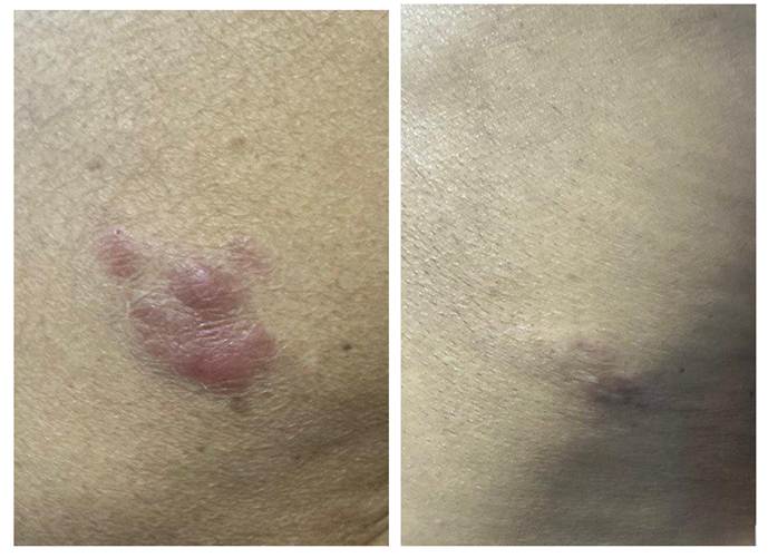
Complete haemogram showed Haemoglobin 10.3 gm/dl, total white blood cell count of 14,000 cells /mm3 , ESR 40 mm/1 hour. She did not test positive for HIV. She underwent an orbital biopsy which revealed caseating granulomas with necrosis, and fragments of destroyed bone however Ziehl-Neelsen staining was negative. PAS & GMS stains for fungus were also negative. Sputum for acid- fast bacilli (AFB) was negative. At one week follow-up the right eyelid wound was healing well, but a soft fluctuant lesion was noted in left eye lower eyelid and aspirated material was sent for Gram’s and Ziehl-Neelsen (ZN) staining, aerobic and AFB culture. She had also developed a soft fluctuant lesion at right lateral malleolus which was also aspirated and sent for staining, aerobic and AFB culture. The Ziehl- Neelsen staining of pus smear from both sites was negative, however at 2 weeks growth was seen on L-J medium on both samples. A smear was prepared from the white colonies cultured on Löwenstein-Jensen (LJ) medium, and stained with ZN stain utilizing a 20% sulfuric acid (H₂SO₄) solution. The microscopic examination revealed the presence of acid-fast bacilli. Subsequently, the growth underwent evaluation through a rapid immunochromatographic diagnostic test known as the MPT 64 antigen detection assay, aimed at distinguishing between Mycobacterium tuberculosis (MTB) and slow growing nontuberculous mycobacteria (NTM). The results of the MPT 64 test demonstrated a positive outcome specifically for Mycobacterium tuberculosis, thereby conclusively affirming the identification of the organism cultivated on Löwenstein-Jensen medium as Mycobacterium tuberculosis 2. A diagnosis of extra-pulmonary orbital tuberculosis was confirmed. She was started on Category 1 Anti tubercular therapy (ATT) consisting of isoniazid (H) 225 mg, rifampicin(R) 225mg, pyrazinamide (Z) 750 mg, ethambutol (E) 600 mg on alternate day were started.
Based on the 2020 WHO Global Tuberculosis report, there were 10 million reported cases of Tuberculosis in 2019, with India contributing to 26 % of the global TB burden. The worldwide incidence of extrapulmonary tuberculosis in 2019 was 16%.3 Not much has changed in the latest WHO global Tuberculosis report which mentions 10.6 million reported cases of tuberculosis in 2022, of which India accounts for 27%.4 The prevalence of extrapulmonary tuberculosis has increased with the emergence of HIV infection, being observed in over 50% of individuals with concurrent TB and AIDS.5, 6 Nevertheless, there are currently no documented cases linking Orbital Tuberculosis (OTB) with HIV or AIDS. The first documented case of orbital tuberculosis dates back to 1881 by Abadie, who illustrated orbital tuberculosis of the lacrimal gland. 7
Orbital tuberculosis can be categorized as primary or secondary. Primary orbital tuberculosis refers to an infection in the ocular area with signs and symptoms confined to that location in absence of systemic involvement. On the other hand, secondary orbital tuberculosis occurs when ocular regions are affected either through hematogenous spread from a distant site or extension from nearby structures, such as the paranasal sinuses. Orbital tuberculosis can manifest as tubercular periostitis or as space-occupying lesions.
Classifying orbital tuberculosis based on site involvement reveals five types 7: (i) classical orbital periostitis (most common), (ii) orbital soft tissue tuberculoma without bony destruction, (iii) orbital tuberculosis with bony involvement; not in category (i), (iv) spread from paranasal sinuses, and (v) dacryoadenitis /adnexal involvement. The case discussed here falls into type 3, where bony involvement was evident in the paranasal sinuses on a CT scan.
Diagnosis relies on history, detecting acid-fast bacilli in eye smears or excised tissue. Given the paucibacillary nature, smear demonstration may be challenging. Clinical suspicion is crucial, and the presence of three key features supports the diagnosis: suggestive clinical presentation, exclusion of other causes, histopathological examination of tissue revealing inflammatory caseating granulomatous lesions, along with a positive culture for Mycobacterium tuberculosis if specimens are collected, response to anti-tubercular treatment, and a present or past history of tuberculosis. In cases where the biopsy is suggestive of caseating granulomatous inflammation but not positive for acid-fast bacilli in the smear, ancillary tests can provide corroborative evidence. It is known that ZN staining has relatively low sensitivity for detecting AFB in histopthologic tissue sections 2. In this scenario, the decision to procure aspirated material for microbiologic evaluation from other affected sites proved conclusive. The gold standard for diagnosis is culture of MTB. When grown on Löwenstein-Jensen media, MTB appears as white (rough buff tough) granular colonies and needs to be incubated for about four weeks due to the slow doubling time (15-20 hours) of MTB. It has been demonstrated that the MPT 64 antigen is exclusively present in viable and actively dividing cells of Mycobacterium tuberculosis.8 The rapid test kit for MPT 64 antigen has exhibited a sensitivity of 99.2%. Notably, a study observed no false positive results, highlighting the reliability of the rapid kit in MTB diagnosis. 9
All patients of orbital/ ocular tuberculosis are to be investigated for AFB in sputum and a chest radiograph should be done. 10 Systemic infection with synchronous clinical evidence is not an uncommon occurrence, albeit smear-positive pulmonary disease remains infrequent. 11 In this child, a fall from the bed, although incidental, appears to have exacerbated the hematoma due to the pre-existing orbital wall erosion and inflammation.
Typically, the diagnosis is established through orbital biopsy, revealing histopathological hallmark of epitheloid granuloma, giant cells and caseating necrosis and/or conclusive microbiological proof of infection. Successful treatment often involves oral antituberculous chemotherapy (ATT), with or without surgery.
Orbital Tuberculosis treatment follows category I and category III of the RNTCP regime, with potential corticosteroid support. Delayed initiation of treatment can have detrimental effects. Hence, maintaining a high index of suspicion is vital for early diagnosis, especially in cases with non-specific symptoms, to prevent long-term complications.
In conclusion, while orbital tuberculosis is exceptionally rare, it should be considered as a differential diagnosis for atypical orbital lesions or non-healing ulcers. Careful evaluation with high suspicion index for infection and emphasis on sending material for not only histopathologic but also microbiologic examination is the cornerstone in obtaining a diagnosis & initiating prompt treatment in order to reduce morbidity and ensure a favourable prognosis. Delayed diagnosis and disease sequelae can significantly impact the patient's quality of life.
Financial support: This research received no specific grant from any public or private funding agency.
Proprietary interest: The authors do not have any financial/proprietary interest in the products or techniques used in this study.
Competing interest statement: No competing interest.
Subscribe now for latest articles and news.