Journal of Medical Sciences and Health
DOI: 10.46347/jmsh.v12.i3.26.134
Year: 2026, Volume: 12, Issue: 3, Pages: 343-346
Case Report
Aastha Sharma 1, Anita Harsh 2
1Senior resident, Department of Pathology, SMS Medical College, Jaipur, Rajasthan, India.
2Senior professor, Department of Pathology, SMS Medical College, Jaipur, Rajasthan, India.
Address for correspondence: Aastha Sharma, Senior resident, Department of Pathology, SMS Medical College, Jaipur, Rajasthan, India.
E-mail: [email protected]
Received Date:15 April 2026, Accepted Date:29 June 2026, Published Date:19 August 2026

Prostate cancer is one of the most common malignancies affecting men worldwide and represents a major cause of cancer-related morbidity and mortality. Although conventional acinar adenocarcinoma accounts for the vast majority of cases, several rare histological variants have been described, each demonstrating distinct morphological, clinical, and prognostic characteristics. Pleomorphic giant cell carcinoma of the prostate (PGCC) is an exceptionally rare and aggressive variant that has recently been recognized in the 2022 World Health Organization (WHO) Classification of Tumours of the Urinary System as a subtype of acinar adenocarcinoma. Histologically, PGCC is characterized by the presence of markedly pleomorphic, bizarre multinucleated giant cells with significant nuclear atypia and abundant cytoplasm. This variant is frequently associated with high-grade prostatic adenocarcinoma and demonstrates aggressive biological behavior with poor clinical outcomes. PGCC most commonly occurs in patients with a prior history of prostate cancer, particularly those who have received androgen deprivation therapy or radiotherapy. Due to its rarity and unusual morphology, diagnosis can be challenging and requires careful clinicopathological correlation along with immunohistochemical analysis. We report a case of PGCC in a 69-year-old male who presented with lower urinary tract symptoms and markedly elevated prostate-specific antigen levels. Histopathological examination combined with immunohistochemistry confirmed the diagnosis. Despite management, the patient succumbed to the disease within a few months, highlighting the aggressive nature of this entity.
Prostate cancer is among the most frequently diagnosed malignancies in men and remains a leading cause of cancer-related deaths globally[1]. The majority of prostate cancers are conventional acinar adenocarcinomas; however, several rare histological variants have been identified, each with unique pathological and clinical features that may influence management and prognosis.
Pleomorphic giant cell carcinoma of the prostate (PGCC) is an extremely rare and poorly characterized variant of prostatic carcinoma. It has recently been incorporated into the 2022 WHO Classification of Tumours of the Urinary System and Male Genital Organs as a subtype of acinar adenocarcinoma[2, 3]. This tumor is defined by the presence of bizarre pleomorphic giant cells showing
marked nuclear atypia and abundant cytoplasm on histopathological examination.
PGCC is considered a highly aggressive neoplasm associated with rapid progression and poor prognosis despite therapeutic interventions[4]. In most cases, this variant coexists with a component of conventional high-grade prostatic adenocarcinoma, suggesting a possible transformation from an existing tumor[5].
A notable feature of PGCC is its frequent occurrence in patients with a prior history of prostate cancer, especially those who have undergone hormonal or radiation therapy. However, it may also arise in advanced untreated or metastatic disease.
A significant diagnostic challenge arises because pleomorphic giant cell carcinomas can occur in multiple organs such as the lung, pancreas, and urinary bladder. Therefore, establishing the primary site is crucial. Immunohistochemical markers including PSA, PSMA, NKX3.1, and androgen receptor are often employed to confirm prostatic origin[2]. Clinical history of prior prostate malignancy further aids in diagnosis[4].
Given the rarity of this tumor and limited data in the literature, reporting individual cases is essential to enhance understanding of its clinicopathological features.
A 69-year-old male presented to the Department of Urology at SMS Medical Hospital, Jaipur, Rajasthan, in December 2024 with complaints of increased frequency of micturition, urgency, hesitancy, and weak urinary stream. These symptoms had progressively worsened over several months. More recently, the patient reported episodes of hematuria and urinary incontinence, along with low-grade back pain and intermittent constipation.
Digital rectal examination revealed a firm, enlarged prostate with multiple discrete nodules corresponding to grade 3 prostatomegaly. The irregular nodular surface raised a strong suspicion of malignancy.
The patient had a known history of carcinoma prostate and had previously received androgen deprivation therapy and radiotherapy at another institution, although prior treatment records were unavailable.
Laboratory investigations showed anemia and mild leukocytosis. Serum PSA level was markedly elevated at 231.3 ng/L. Pelvic MRI demonstrated a large prostatic mass measuring approximately 82 × 80 × 78 mm with areas of cystic degeneration. The mass exerted pressure on adjacent structures including the urinary bladder and rectum but showed no definite invasion or pelvic lymphadenopathy.
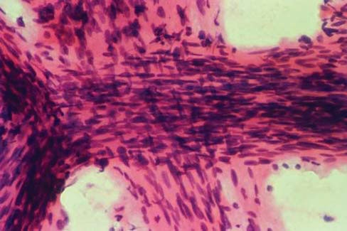
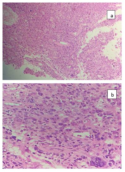
A transrectal prostate biopsy was performed. Tissue fragments were fixed in 10% formalin and processed routinely. Hematoxylin and eosin-stained sections revealed tumor composed of a mixture of round, oval, spindle-shaped, and markedly pleomorphic giant cells. These cells exhibited abundant eosinophilic and occasionally vacuolated cytoplasm. The nuclei were highly pleomorphic, hyperchromatic, and often lobulated with prominent nucleoli. Multinucleation was a prominent feature, contributing to the striking pleomorphic appearance ([Fig. 1] a,b).
Additional findings included areas of coagulative necrosis, hemorrhage with fibrin deposition, and dense acute inflammatory infiltrates. No lymphovascular or perineural invasion was identified.
Based on these morphological features, a diagnosis of pleomorphic giant cell carcinoma of the prostate was made.
Immunohistochemical analysis showed tumor cells positive for Pan-cytokeratin (PanCK) and AMACR, while negative for GATA-3 and PSA ([Fig. 2] a,b).
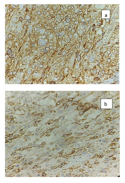
These findings supported prostatic epithelial origin and helped exclude urothelial carcinoma.
Despite supportive care and treatment, the patient’s condition deteriorated rapidly, and he succumbed to the disease in April 2025.
Pleomorphic giant cell carcinoma is a rare tumor type described in several organs including the hepatobiliary system, pancreas, thyroid, urinary bladder, kidney, and endometrium[3, 5]. In the prostate, it was first reported by Mai et al. in 1996[7]. Since then, only a limited number of cases have been documented. This variant is clinically significant due to its aggressive nature and poor prognosis. Patients often present with advanced disease and have significantly shorter survival compared to those with conventional adenocarcinoma[8].
Histologically, PGCC is characterized by large, discohesive tumor cells with marked pleomorphism, prominent nucleoli, and frequent multinucleation. Cytoplasm may contain hyaline inclusions, and atypical mitotic figures are common. Necrosis is frequently observed. Unlike sarcomatoid carcinoma, PGCC typically lacks a dominant spindle cell component. The proportion of pleomorphic giant cells varies widely, ranging from 5% to 70% of the tumor[9].
The pleomorphic component may arise in association with or as a transformation of high-grade adenocarcinoma or other epithelial tumors of the prostate[10]. According to WHO guidelines, Gleason grading is not applied to PGCC; however, any associated conventional adenocarcinoma component should be graded separately. Most cases show high-grade features corresponding to Grade Group 5[11].
The exact pathogenesis remains unclear. Some studies suggest a role of prior therapy such as radiotherapy or chemotherapy in inducing this morphology, although evidence is inconclusive[12].
Differential diagnoses include radiation-induced atypia, sarcomatoid carcinoma, urothelial carcinoma involving the prostate, and metastatic carcinoma. Radiation atypia shows degenerative changes rather than true malignancy. Sarcomatoid carcinoma demonstrates a prominent spindle cell component. Urothelial carcinoma can be identified by in situ lesions and immunohistochemical markers. Metastatic tumors require clinical and pathological correlation[11].
Alharbi et al[1]. reported 30 cases of prostatic adenocarcinoma with pleomorphic giant cell features, most presenting with advanced disease and poor survival.
Bilé-Silva et al.[4] analyzed 20 cases, noting variable proportions of giant cells and heterogeneous PSA expression.
Parwani et al.[5] described cases where PGCC coexisted with other carcinoma types such as squamous, small cell, and ductal carcinoma.
Pleomorphic giant cell carcinoma of the prostate is a rare but highly aggressive variant of prostatic carcinoma. It is commonly associated with high-grade disease and may develop following prior therapy. The tumor demonstrates rapid progression and poor clinical outcomes.
Accurate recognition is essential due to its diagnostic challenges and prognostic implications. Histopathological examination combined with immunohistochemistry plays a crucial role in diagnosis. Continued reporting of such cases is necessary to better understand its biological behavior and guide future therapeutic strategies[4].
1. Cancer Publications in One Year (2022): A Cross-Sectional Study. Barw Medical Journal. 2023; 1 (2). Available from: https://doi.org/10.58742/bmj.v1i2.30
2. The 2022 World Health Organization Classification of Tumours of the Urinary System and Male Genital Organs—Part A: Renal, Penile, and Testicular Tumours. European Urology. 2022; 82 (5). Available from: https://doi.org/10.1016/j.eururo.2022.06.016
3. Pleomorphic giant cell carcinoma of prostate: Rare tumor with unique clinicopathological, immunohistochemical, and molecular features. Annals of Diagnostic Pathology. 2021; 52 Available from: https://doi.org/10.1016/j.anndiagpath.2021.151719
4. Pleomorphic giant cell carcinoma of the prostate: clinicopathologic analysis and oncological outcomes. Virchows Archiv. 2023; 482 (3). Available from: https://doi.org/10.1007/s00428-022-03481-7
5. Pleomorphic Giant Cell Adenocarcinoma of the Prostate. American Journal of Surgical Pathology. 2006; 30 (10). Available from: https://doi.org/10.1097/01.pas.0000209841.77595.4b
6. Pleomorphic Giant Cell Carcinoma of the Prostate. Archives of Pathology & Laboratory Medicine. 2005; 129 (5). Available from: https://doi.org/10.5858/2005-129-0683-pgccot
7. Mai K, Burns B, Morash D. Giant‑cell carcinoma of the prostate. Journal of Urologic Pathology 5: 167‑174, 1996.
8. Moch H, Humphrey PA, Ulbright TM, Reuter VE. WHO Classification of Tumours of the Urinary Systemandth Male Genital Organs. 4 ed. Lyon: IARC Press. 2016.
9. Zynger DL, Parwani AV. Acinar adenocarcinoma, variant differentiation, Chapter 2: Premalignant conditions and prostate carcinoma In: Prostate Pathology: DSPG, Demos Medical Publishing LLC, New York 2015; 126-1288.
10. Amin MB, Tickoo SK. Pleomorphic giant cell adenocarcinoma. In: Diagnostic Pathologynd Genitourinary, 2 ed., Elsevier, Philadelphia, 2016: 628
11. Li J, Wang Z. The pathology of unusual subtypes of prostate cancer. Chinese Journal of Cancer Research 2016;28(1):130-143.
12. Carcinosarcoma and sarcomatoid carcinoma of the bladder: clinicopathological study of 41 cases. Journal of Urology. 1998; 159 (5). Available from: https://doi.org/10.1097/00005392-199805000-00023
Subscribe now for latest articles and news.